. 2024 Feb 26:15:1335998.
doi: 10.3389/fimmu.2024.1335998. eCollection 2024.

Bi-specific autoantigen-T cell engagers as targeted immunotherapy for autoreactive B cell depletion in autoimmune diseases

Affiliations

Bi-specific autoantigen-T cell engagers as targeted immunotherapy for autoreactive B cell depletion in autoimmune diseases

Luca Perico et al. Front Immunol. .

Erratum in

Abstract

Introduction: In autoimmune diseases, autoreactive B cells comprise only the 0.1-0.5% of total circulating B cells. However, current first-line treatments rely on non-specific and general suppression of the immune system, exposing patients to severe side effects. For this reason, identification of targeted therapies for autoimmune diseases is an unmet clinical need.

Methods: Here, we designed a novel class of immunotherapeutic molecules, Bi-specific AutoAntigen-T cell Engagers (BiAATEs), as a potential approach for targeting the small subset of autoreactive B cells. To test this approach, we focused on a prototype autoimmune disease of the kidney, membranous nephropathy (MN), in which phospholipase A2 receptor (PLA2R) serves as primary nephritogenic antigen. Specifically, we developed a BiAATE consisting of the immunodominant Cysteine-Rich (CysR) domain of PLA2R and the single-chain variable fragment (scFv) of an antibody against the T cell antigen CD3, connected by a small flexible linker.

Results: BiAATE creates an immunological synapse between autoreactive B cells bearing an CysR-specific surface Ig+ and T cells. Ex vivo, the BiAATE successfully induced T cell-dependent depletion of PLA2R-specific B cells isolated form MN patients, sparing normal B cells. Systemic administration of BiAATE to mice transgenic for human CD3 reduced anti-PLA2R antibody levels following active immunization with PLA2R.

Discussion: Should this approach be confirmed for other autoimmune diseases, BiAATEs could represent a promising off-the-shelf therapy for precision medicine in virtually all antibody-mediated autoimmune diseases for which the pathogenic autoantigen is known, leading to a paradigm shift in the treatment of these diseases.

Keywords: anti-PLA2R antibodies; autoimmune diseases; autoreactive B cell; bi-specific autoantigen-T cell engagers; membranous nephropathy; targeted immunotherapies.


Conflict of interest statement

FS, PI-P and KT were employed by genOway. The remaining authors declare that the research was conducted in the absence of any commercial or financial relationships that could be construed as a potential conflict of interest. The author(s) declared that they were an editorial board member of Frontiers, at the time of submission. This had no impact on the peer review process and the final decision.

Figures
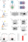
Figure 1
Figure 1
Design and characterization of the BiAATE for MN. (A, B) Schematic representation of the structure (A) and the proposed mechanism of action (B) of the BiAATE, a bi-specific molecule that expresses the autoantigen involved in a specific autoimmune disease linked to the single-chain variable fragment (scFv) of an anti-CD3 by a small flexible linker. As such, the BiAATE redirects the lytic activity of T cells against the surface immunoglobulin (sIg)+ included in the B cell receptor (BCR) of autoreactive B cells. Activation of T cells leads to the production of perforin (PFN), granzyme B (GzmB), interferon γ (IFNγ), and tumor necrosis factor α (TNFα) that induce the selective removal of autoreactive B cells. All drawings were created by using BioRender.com. (C) Representative ponceau red staining of 1 μg BiAATE loaded under reducing (R) and non-reducing (NR) condition. Molecular weights (MW) are reported for each Western Blot and expressed in kilo Dalton (kDa). The BiAATE is indicated by arrows and exhibited a MW of approximatively 50 kDa. (D) Schematic representation of the Western Blot performed to test the immunogenic properties of the CysR antigen included in the BiAATE. (E) Representative Western Blot of 1 μg BiAATE incubated with sera from healthy controls (CTRL sera) or MN patients (MN sera). Data derive from n=6 experiments from three independent batches of BiAATE. (F) Schematic representation of the Western Blot performed to test the ability of the BiAATE to simultaneously bind CD3 in T cells and the pathogenic antibodies in the MN sera. (G) Representative Western Blot of total peripheral blood mononuclear cell (PBMC) extracts loaded under R and NR condition and incubated with 5 μg/mL BiAATE followed by exposure to CTRL sera (BiAATE + CTRL sera) or MN sera (BiAATE + MN sera), or incubated with MN sera without prior exposure to 5 μg/mL BiAATE (MN sera). Actin was used as sample loading control. Representative western blot of CD3 expression in PBMC extracts to confirm CD3 specificity (anti-human CD3). Data derive from n=6 experiments with PBMCs isolated from 6 healthy subjects and exposed to sera from n=6 CTRL or MN patients. (H) Representative dot plots of anti-human IgG4 FITC antibody binding and 7AAD staining after gating singlet lymphocytes. PBMCs were incubated with 5 µg/mL BiAATEs in the presence of CTRL or MN sera, then with mouse anti-human IgG4 FITC antibody and 7AAD. (I) Percentage of lymphocytes bound by anti-human IgG4 FITC antibody after incubation with 5 µg/mL BiAATEs in the presence of CTRL or MN sera (n=6 per group). Data represent mean ± SEM and were analyzed using unpaired t-test. **p-value<0.01 vs CTRL sera.
Figure 2
Figure 2
The BiAATE selectively depletes PLA2R-specific autoreactive B cells isolated from MN patients. (A, B) Schematic representation of the experimental procedure (A) and quantification overtime of IgG anti-PLA2R in the supernatants (B) during the expansion of B cells isolated from CTRL and MN PBMCs treated or not with 3 μg/mL BiAATE for 15 hours (n=4 per group). Data represent mean ± SEM and were analyzed using 2-way ANOVA corrected with Tukey post hoc test. *p-value<0.05, and **p-value<0.01 vs MN at 14 days; °°p-value<0.01 vs CTRL at the corresponding time; ## p-value<0.01 vs CTRL + BiAATE at the corresponding time; and $p-value<0.05 vs MN+ BiAATE at the corresponding time. (C, D) Schematic representation of the experimental procedure (C) and quantification overtime of IgG anti-PLA2R antibody in the supernatants (D) during the expansion of B cells isolated from PBMCs of MN patients (n=6 per group). Data represent mean ± SEM and were analyzed using 1-way ANOVA corrected with Tukey post hoc test. *p-value<0.05 vs 14 days; °°p-value<0.01 vs 10 days. (E) Quantification of IgG anti-PLA2R in the supernatants of expanded B cells from MN patients treated with autologous T cells alone or in the presence of 3 μg/mL BiAATEs or 3 μg/mL Blinatumomab (BLINA) for 3 days (n=6 per group). Data represent mean ± SEM and were analyzed using 1-way ANOVA corrected with Tukey post hoc test. *p-value<0.05, **p-value<0.01, and ***p-value<0.001 vs T cells alone. (F) Percentages on singlets of viable (7AAD negative) CD3-CD19+ B cells co-cultured with autologous T cells alone or in the presence of 3 μg/mL BiAATE or 3 μg/mL BLINA for 3 days (n=6 per group). The percentage of viable B cells in co-culture with autologous T cells alone were taken as 100%. Data represent mean ± SEM and were analyzed using 1-way ANOVA corrected with Tukey post hoc test. ***p-value<0.001 vs T cells alone and °°°p-value<0.001 vs BIAATE.
Figure 3
Figure 3
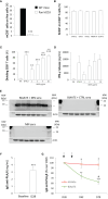
Human CD3 is expressed and functional on Pan hCD3 mice, and the BiAATE reduces PLA2R antibody titer in these mice following active immunization with PLA2R. (A) Expression of mouse CD3 in splenocytes from Pan hCD3 and WT mice (n=3) determined flow cytometry. Data represent mean ± SEM and were analyzed using unpaired t-test. ***p-value<0.001 vs WT mice. (B) Binding of different antibody clones of human anti-CD3 (SP34.2, OKT3, HIT3α, MEM-57, UCHT-1) to CD4+ T cells from Pan hCD3 mice (n=3) determined by flow cytometry. (C, D) Percentage of dividing CD8+ T cells (C) and IFN-γ release (D) determined by flow cytometry and ELISA, respectively, in unstimulated (NA) T cells isolated from Pan hCD3 or in response to coated human anti-CD3 (OKT3) activation for 3 days at different concentration (n=6 per group). Human anti-CD3 antibody clone SP34.2 (5 µg/mL) was used as a positive control. Data represent mean ± SEM and were analyzed using 1-way ANOVA corrected with Tukey post hoc test. **p-value<0.01, and ***p-value<0.001 vs NA; °°°p-value<0.001 vs OKT3 1 μg/mL. (E) Representative Western Blot of MN patients’ IgG4 binding to CD3 in total splenocyte extracts from hCD3 mice (n=6) incubated with (upper panel on the left) or without (lower panel on the left) 5 μg/mL BiAATE followed by MN sera. The right panel shows representative Western Blot of IgG4 binding to CD3 in total splenocyte extracts from hCD3 mice (n=6) incubated with 5 μg/mL BiAATE followed by incubation with CTRL sera. Actin was used as sample loading control. MW are reported in each Western Blot and expressed in kDa. (F) Quantification of IgG anti-PLA2R (mg/mL) in the sera of pan hCD3 mice (n=18) immunized with two doses of 50 μg PLA2R antigen. Data represent mean ± SEM and were analyzed with unpaired t-test. ***p-value<0.001 vs Baseline. (G) Quantification overtime of IgG anti-PLA2R expressed as % changes over D28 in the sera of immunized pan hCD3 mice (n=9 per group) treated at D28 and D35 (black arrows) with vehicle or 1 mg/kg BiAATE. Data represent mean ± SEM and were analyzed with 2-way ANOVA corrected with Šídák’s multiple comparisons test. *p-value<0.05 vs BiAATE at the corresponding time; °°p-value<0.01, and °°°p-value<0.001 vs BiAATE at D28; and #p-value<0.05 vs BiAATE at D42.

References

    1. Walsh SJ, Rau LM. Autoimmune diseases: a leading cause of death among young and middle-aged women in the United States. Am J Public Health (2000) 90:1463–6. doi: 10.2105/ajph.90.9.1463 - DOI - PMC - PubMed
    1. Scolari F, Alberici F, Mescia F, Delbarba E, Trujillo H, Praga M, et al. . Therapies for membranous nephropathy: A tale from the old and new millennia. Front Immunol (2022) 13:789713. doi: 10.3389/fimmu.2022.789713 - DOI - PMC - PubMed
    1. Minard-Colin V, Aupérin A, Pillon M, Burke GAA, Barkauskas DA, Wheatley K, et al. . Rituximab for high-risk, mature B-cell non-hodgkin’s lymphoma in children. N Engl J Med (2020) 382:2207–19. doi: 10.1056/NEJMoa1915315 - DOI - PMC - PubMed
    1. Remuzzi G, Chiurchiu C, Abbate M, Brusegan V, Bontempelli M, Ruggenenti P. Rituximab for idiopathic membranous nephropathy. Lancet (2002) 360:923–4. doi: 10.1016/S0140-6736(02)11042-7 - DOI - PubMed
    1. Ruggenenti P, Fervenza FC, Remuzzi G. Treatment of membranous nephropathy: time for a paradigm shift. Nat Rev Nephrol (2017) 13:563–79. doi: 10.1038/nrneph.2017.92 - DOI - PubMed

Publication types

LinkOut - more resources