

The Mla system and its role in maintaining outer membrane barrier function in Stenotrophomonas maltophilia
- PMID: 38469346
- PMCID: PMC10925693
- DOI: 10.3389/fcimb.2024.1346565
The Mla system and its role in maintaining outer membrane barrier function in Stenotrophomonas maltophilia
Abstract
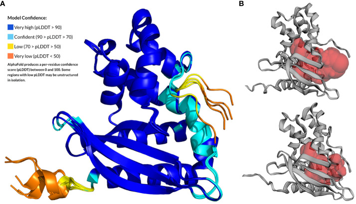
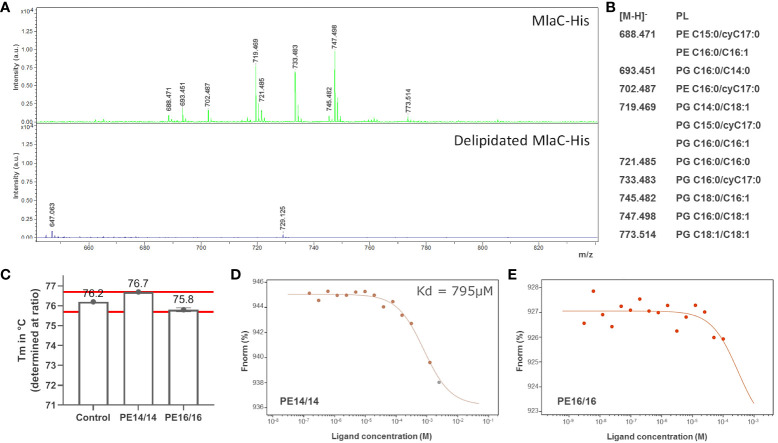
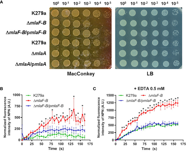
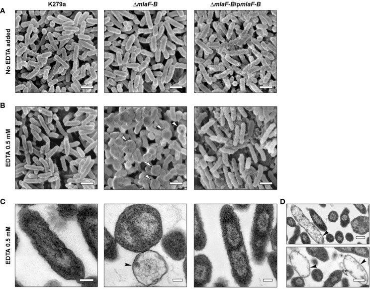
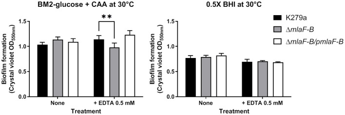
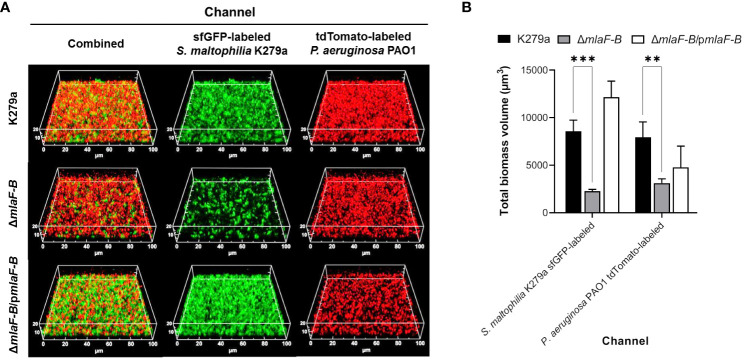
Stenotrophomonas maltophilia are ubiquitous Gram-negative bacteria found in both natural and clinical environments. It is a remarkably adaptable species capable of thriving in various environments, thanks to the plasticity of its genome and a diverse array of genes that encode a wide range of functions. Among these functions, one notable trait is its remarkable ability to resist various antimicrobial agents, primarily through mechanisms that regulate the diffusion across cell membranes. We have investigated the Mla ABC transport system of S. maltophilia, which in other Gram-negative bacteria is known to transport phospholipids across the periplasm and is involved in maintaining outer membrane homeostasis. First, we structurally and functionally characterized the periplasmic substrate-binding protein MlaC, which determines the specificity of this system. The predicted structure of the S. maltophilia MlaC protein revealed a hydrophobic cavity of sufficient size to accommodate the phospholipids commonly found in this species. Moreover, recombinant MlaC produced heterologously demonstrated the ability to bind phospholipids. Gene knockout experiments in S. maltophilia K279a revealed that the Mla system is involved in baseline resistance to antimicrobial and antibiofilm agents, especially those with divalent-cation chelating activity. Co-culture experiments with Pseudomonas aeruginosa also showed a significant contribution of this system to the cooperation between both species in the formation of polymicrobial biofilms. As suggested for other Gram-negative pathogenic microorganisms, this system emerges as an appealing target for potential combined antimicrobial therapies.
Keywords: Mla system; Stenotrophomonas maltophilia; biofilm; chelating agents; membrane permeability.
Copyright © 2024 Coves, Mamat, Conchillo-Solé, Huedo, Bravo, Gómez, Krohn, Streit, Schaible, Gibert, Daura and Yero.
Conflict of interest statement
The authors declare that the research was conducted in the absence of any commercial or financial relationships that could be construed as a potential conflict of interest. The author(s) declared that they were an editorial board member of Frontiers, at the time of submission. This had no impact on the peer review process and the final decision.
Figures
Similar articles
-
Molecular Insight into Gene Response of Diorcinol- and Rubrolide-Treated Biofilms of the Emerging Pathogen Stenotrophomonas maltophilia.Microbiol Spectr. 2022 Jun 29;10(3):e0258221. doi: 10.1128/spectrum.02582-21. Epub 2022 Apr 26. Microbiol Spectr. 2022. PMID: 35471093 Free PMC article.
-
Cooperativity between Stenotrophomonas maltophilia and Pseudomonas aeruginosa during Polymicrobial Airway Infections.Infect Immun. 2020 Mar 23;88(4):e00855-19. doi: 10.1128/IAI.00855-19. Print 2020 Mar 23. Infect Immun. 2020. PMID: 31932329 Free PMC article.
-
The rpf/DSF signalling system of Stenotrophomonas maltophilia positively regulates biofilm formation, production of virulence-associated factors and β-lactamase induction.FEMS Microbiol Lett. 2019 Mar 1;366(6):fnz069. doi: 10.1093/femsle/fnz069. FEMS Microbiol Lett. 2019. PMID: 31044250
-
Stenotrophomonas maltophilia biofilm: its role in infectious diseases.Expert Rev Anti Infect Ther. 2019 Nov;17(11):877-893. doi: 10.1080/14787210.2019.1685875. Epub 2019 Nov 1. Expert Rev Anti Infect Ther. 2019. PMID: 31658838 Review.
-
Stenotrophomonas maltophilia - a low-grade pathogen with numerous virulence factors.Infect Dis (Lond). 2019 Mar;51(3):168-178. doi: 10.1080/23744235.2018.1531145. Epub 2018 Nov 13. Infect Dis (Lond). 2019. PMID: 30422737 Review.
Cited by
-
A chimeric Mla-Pqi lipid transport system is required for Brucella abortus survival in macrophages.EMBO J. 2025 Aug 13. doi: 10.1038/s44318-025-00511-3. Online ahead of print. EMBO J. 2025. PMID: 40804184
-
Breaking Barriers: Exploiting Envelope Biogenesis and Stress Responses to Develop Novel Antimicrobial Strategies in Gram-Negative Bacteria.Pathogens. 2024 Oct 11;13(10):889. doi: 10.3390/pathogens13100889. Pathogens. 2024. PMID: 39452760 Free PMC article. Review.
-
An acyl-homoserine lactone acylase found in Stenotrophomonas maltophilia exhibits both quorum quenching activity and the ability to degrade penicillin antibiotics.Sci Rep. 2025 Mar 12;15(1):8557. doi: 10.1038/s41598-025-92749-4. Sci Rep. 2025. PMID: 40074792 Free PMC article.
-
RamA upregulates the ATP-binding cassette transporter mlaFEDCB to mediate resistance to tetracycline-class antibiotics and the stability of membranes in Klebsiella pneumoniae.Microbiol Spectr. 2025 Feb 4;13(2):e0172824. doi: 10.1128/spectrum.01728-24. Epub 2024 Dec 31. Microbiol Spectr. 2025. PMID: 39745369 Free PMC article.
References
-
- Alcaraz E., García C., Friedman L., de Rossi B. P. (2019). The rpf/DSF signalling system of Stenotrophomonas maltophilia positively regulates biofilm formation, production of virulence-associated factors and β-lactamase induction. FEMS Microbiol. Lett. 366, fnz069. doi: 10.1093/femsle/fnz069 - DOI - PubMed
-
- Alio I., Gudzuhn M., Pérez García P., Danso D., Schoelmerich M. C., Mamat U., et al. . (2020). Phenotypic and transcriptomic analyses of seven clinical Stenotrophomonas maltophilia isolates identify a small set of shared and commonly regulated genes involved in the biofilm lifestyle. Appl. Environ. Microbiol. 86, e02038–e02020. doi: 10.1128/AEM.02038-20 - DOI - PMC - PubMed
-
- Alio I., Moll R., Hoffmann T., Mamat U., Schaible U. E., Pappenfort K., et al. . (2023). Stenotrophomonas maltophilia affects the gene expression profiles of the major pathogens Pseudomonas aeruginosa and Staphylococcus aureus in an in vitro multispecies biofilm model. Microbiol. Spectr. 11, e0085923. doi: 10.1128/spectrum.00859-23 - DOI - PMC - PubMed
MeSH terms
Substances
LinkOut - more resources
Full Text Sources
Molecular Biology Databases
Research Materials