

Locus coeruleus features are linked to vagus nerve stimulation response in drug-resistant epilepsy
- PMID: 38469571
- PMCID: PMC10926962
- DOI: 10.3389/fnins.2024.1296161
Locus coeruleus features are linked to vagus nerve stimulation response in drug-resistant epilepsy
Abstract
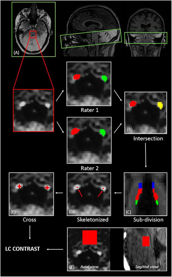
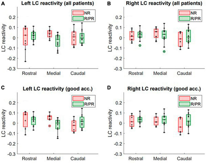
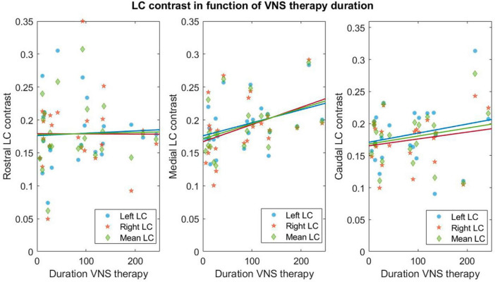
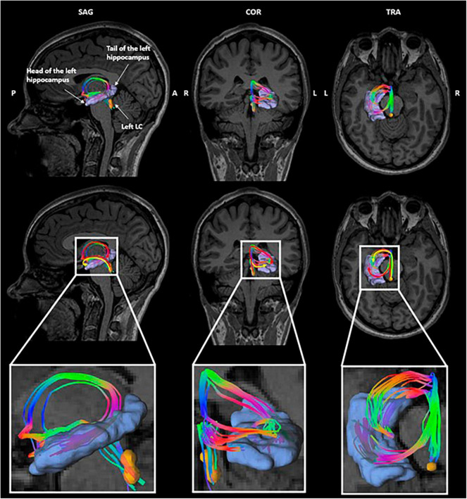
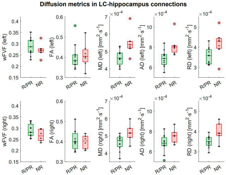
The locus coeruleus-norepinephrine system is thought to be involved in the clinical effects of vagus nerve stimulation. This system is known to prevent seizure development and induce long-term plastic changes, particularly with the release of norepinephrine in the hippocampus. However, the requisites to become responder to the therapy and the mechanisms of action are still under investigation. Using MRI, we assessed the structural and functional characteristics of the locus coeruleus and microstructural properties of locus coeruleus-hippocampus white matter tracts in patients with drug-resistant epilepsy responding or not to the therapy. Twenty-three drug-resistant epileptic patients with cervical vagus nerve stimulation were recruited for this pilot study, including 13 responders or partial responders and 10 non-responders. A dedicated structural MRI acquisition allowed in vivo localization of the locus coeruleus and computation of its contrast (an accepted marker of LC integrity). Locus coeruleus activity was estimated using functional MRI during an auditory oddball task. Finally, multi-shell diffusion MRI was used to estimate the structural properties of locus coeruleus-hippocampus tracts. These characteristics were compared between responders/partial responders and non-responders and their association with therapy duration was also explored. In patients with a better response to the therapy, trends toward a lower activity and a higher contrast were found in the left medial and right caudal portions of the locus coeruleus, respectively. An increased locus coeruleus contrast, bilaterally over its medial portions, correlated with duration of the treatment. Finally, a higher integrity of locus coeruleus-hippocampus connections was found in patients with a better response to the treatment. These new insights into the neurobiology of vagus nerve stimulation may provide novel markers of the response to the treatment and may reflect neuroplasticity effects occurring in the brain following the implantation.
Keywords: biomarker; epilepsy; locus coeruleus; magnetic resonance imaging; vagus nerve stimulation.
Copyright © 2024 Berger, Beckers, Joris, Duchêne, Danthine, Delinte, Cakiroglu, Sherif, Morrison, Sánchez, Macq, Dricot, Vandewalle and El Tahry.
Conflict of interest statement
AB was an employee of Synergia Medical SA. The firm was not involved in the study design, data collection and analysis, interpretation of the data, the writing of the article, or the decision to submit it for publication. GD was employed by General Electric Healthcare. The remaining authors declare that the research was conducted in the absence of any commercial or financial relationships that could be construed as a potential conflict of interest.
Figures
Similar articles
-
Identifying responders to vagus nerve stimulation based on microstructural features of thalamocortical tracts in drug-resistant epilepsy.Neurotherapeutics. 2024 Sep;21(5):e00422. doi: 10.1016/j.neurot.2024.e00422. Epub 2024 Jul 4. Neurotherapeutics. 2024. PMID: 38964949 Free PMC article.
-
How Is the Norepinephrine System Involved in the Antiepileptic Effects of Vagus Nerve Stimulation?Front Neurosci. 2021 Dec 2;15:790943. doi: 10.3389/fnins.2021.790943. eCollection 2021. Front Neurosci. 2021. PMID: 34924947 Free PMC article. Review.
-
Recordings from the rat locus coeruleus during acute vagal nerve stimulation in the anaesthetised rat.Neurosci Lett. 2005 May 13;379(3):174-9. doi: 10.1016/j.neulet.2004.12.055. Epub 2005 Jan 26. Neurosci Lett. 2005. PMID: 15843058
-
The role of locus coeruleus in the antiepileptic activity induced by vagus nerve stimulation.Eur J Neurosci. 2011 Jun;33(12):2169-78. doi: 10.1111/j.1460-9568.2011.07707.x. Epub 2011 May 3. Eur J Neurosci. 2011. PMID: 21535457 Review.
-
Transcutaneous auricular vagus nerve stimulation at 1 Hz modulates locus coeruleus activity and resting state functional connectivity in patients with migraine: An fMRI study.Neuroimage Clin. 2019;24:101971. doi: 10.1016/j.nicl.2019.101971. Epub 2019 Aug 5. Neuroimage Clin. 2019. PMID: 31648171 Free PMC article. Clinical Trial.
Cited by
-
Attention-dependent coupling with forebrain and brainstem neuromodulatory nuclei differs across the lifespan.Geroscience. 2025 Jun;47(3):4301-4320. doi: 10.1007/s11357-025-01582-0. Epub 2025 Mar 4. Geroscience. 2025. PMID: 40038158 Free PMC article.
-
Transcutaneous auricular vagus nerve stimulation can modulate fronto-parietal brain networks.Front Neurosci. 2024 Jul 18;18:1368754. doi: 10.3389/fnins.2024.1368754. eCollection 2024. Front Neurosci. 2024. PMID: 39091347 Free PMC article.
References
-
- Aston-Jones G., Shipley M. T., Chouvet G., Ennis M., van Bockstaele E., Pieribone V., et al. (1991). Afferent regulation of locus coeruleus neurons: Anatomy, physiology and pharmacology. Prog. Brain Res. 88 47–75. - PubMed
-
- Beck D., de Lange A. M., Maximov I. I., Richard G., Andreassen O. A., Nordvik J. E., et al. (2021). White matter microstructure across the adult lifespan: A mixed longitudinal and cross-sectional study using advanced diffusion models and brain-age prediction. Neuroimage 224:117441. 10.1016/j.neuroimage.2020.117441 - DOI - PubMed
LinkOut - more resources
Full Text Sources