

Retinal structure and vessel density changes in cerebral small vessel disease
- PMID: 38469574
- PMCID: PMC10925719
- DOI: 10.3389/fnins.2024.1288380
Retinal structure and vessel density changes in cerebral small vessel disease
Abstract
Background: Cerebral small vessel disease (CSVD) attaches people's attention in recent years. In this study, we aim to explore retinal structure and vessel density changes in CSVD patients.
Methods: We collected information on retinal metrics assessed by optical coherence tomography (OCT) and OCT angiography and CSVD characters. Logistic and liner regression was used to analyze the relationship between retinal metrics and CSVD.
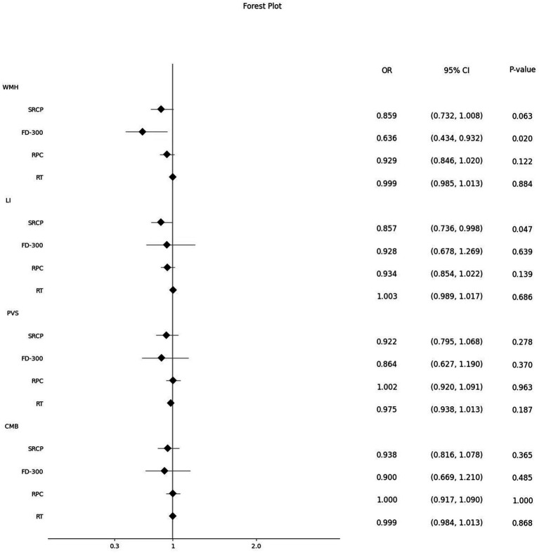
Results: Vessel density of superficial retinal capillary plexus (SRCP), foveal density- 300 length (FD-300), radial peripapillary capillary (RPC) and thickness of retina were significantly lower in CSVD patients, the difference only existed in the thickness of retina after adjusted relevant risk factors (OR (95% CI): 0.954 (0.912, 0.997), p = 0.037). SRCP vessel density showed a significant downward trend with the increase of CSVD scores (β: -0.087, 95%CI: -0.166, -0.008, p = 0.031). SRCP and FD-300 were significantly lower in patients with lacunar infarctions and white matter hypertensions separately [OR (95% CI): 0.857 (0.736, 0.998), p = 0.047 and OR (95% CI): 0.636 (0.434, 0.932), p = 0.020, separately].
Conclusion: SRCP, FD-300 and thickness of retina were associated with the occurrence and severity of total CSVD scores and its different radiological manifestations. Exploring CSVD by observing alterations in retinal metrics has become an optional research direction in future.
Keywords: cerebral small vessel disease (CSVD); diagnosis; neuro-ophtalmology; retinal structure; retinal vessel density.
Copyright © 2024 Wang, Wang, Wang, Du, Wang, Wang, Yang and Zhao.
Conflict of interest statement
The authors declare that the research was conducted in the absence of any commercial or financial relationships that could be construed as a potential conflict of interest.
Figures
Similar articles
-
The vessel density of the superficial retinal capillary plexus as a new biomarker in cerebral small vessel disease: an optical coherence tomography angiography study.Neurol Sci. 2021 Sep;42(9):3615-3624. doi: 10.1007/s10072-021-05038-z. Epub 2021 Jan 11. Neurol Sci. 2021. PMID: 33432462
-
Fundus Changes Evaluated by OCTA in Patients With Cerebral Small Vessel Disease and Their Correlations: A Cross-Sectional Study.Front Neurol. 2022 Apr 25;13:843198. doi: 10.3389/fneur.2022.843198. eCollection 2022. Front Neurol. 2022. PMID: 35547389 Free PMC article.
-
Association between retinal vessel density and neuroimaging features and cognitive impairment in cerebral small vessel disease.Clin Neurol Neurosurg. 2022 Oct;221:107407. doi: 10.1016/j.clineuro.2022.107407. Epub 2022 Aug 3. Clin Neurol Neurosurg. 2022. PMID: 35933965
-
Retinal biomarkers of Cerebral Small Vessel Disease: A systematic review.PLoS One. 2022 Apr 14;17(4):e0266974. doi: 10.1371/journal.pone.0266974. eCollection 2022. PLoS One. 2022. PMID: 35421194 Free PMC article.
-
Increased Extracellular Water in Normal-Appearing White Matter in Patients with Cerebral Small Vessel Disease.J Integr Neurosci. 2024 Feb 22;23(2):46. doi: 10.31083/j.jin2302046. J Integr Neurosci. 2024. PMID: 38419445 Review.
Cited by
-
Retinal vessel density and cognitive function in healthy older adults.Exp Brain Res. 2025 Apr 15;243(5):114. doi: 10.1007/s00221-025-07076-x. Exp Brain Res. 2025. PMID: 40232349 Free PMC article.
References
-
- Albert M. S., DeKosky S. T., Dickson D., Dubois B., Feldman H. H., Fox N. C., et al. . (2011). The diagnosis of mild cognitive impairment due to alzheimer's disease: recommendations from the national institute on aging-alzheimer's association workgroups on diagnostic guidelines for alzheimer's disease. Alzheimers Dement. 7, 270–279. doi: 10.1016/j.jalz.2011.03.008, PMID: - DOI - PMC - PubMed
LinkOut - more resources
Full Text Sources