. 2024 Mar 9:16:91505.
doi: 10.52965/001c.91505. eCollection 2024.

Distal Fibular Metastasis of Colorectal Carcinoma: A Case Report

Distal Fibular Metastasis of Colorectal Carcinoma: A Case Report

Lauren Luther et al. Orthop Rev (Pavia). .

Abstract

Case: A 62-year-old woman presenting with ankle pain was initially treated for a non-displaced fracture. Persistent pain despite months of conservative management for her presumed injury prompted repeat radiographs which demonstrated the progression of a lytic lesion and led to an orthopedic oncology referral. Following a complete work-up, including biopsy and staging, she was diagnosed with colorectal carcinoma metastatic to the distal fibula.

Conclusion: Secondary tumors of the fibula are uncommon but an important diagnosis to consider for intractable lower extremity pain especially in patients with history of malignancy or lack of age-appropriate cancer screening.

Keywords: ankle pain; colon cancer; fibula; lytic lesion; metastatic; tumor.


Conflict of interest statement

There are no disclosures relevant for this article.

Figures

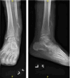
Figure 1.
Figure 1.. AP and lateral radiographs at patient’s initial presentation demonstrate a lytic meta-diaphyseal lesion with erosion of the posterior cortex.
Figure 2.
Figure 2.. Radiographs after re-injury 12-weeks following initial presentation reveal a new pathologic fracture and progression of the fibular lesion.
Figure 3.
Figure 3.. Radiographs demonstrate extensive bone destruction of the distal fibula, an associated soft tissue mass, and diffuse osteopenia.
Figure 4.
Figure 4.. CT chest/abdomen/pelvis with and without contrast. Axial plane images with a red circle showing large circumferential soft tissue mass extending to perineal soft tissues, vaginal cuff, and left gluteal fold (left panel). Large destructive soft tissue mass with significant osseous destruction of the sacrum and coccyx (right panel).
Figure 5.
Figure 5.. CT chest/abdomen/pelvis with and without contrast. Coronal plane images with a red circle showing large circumferential soft tissue mass extending to perineal soft tissues, vaginal cuff, and left gluteal fold (left panel). Large destructive soft tissue mass with significant osseous destruction of the sacrum and coccyx (right panel).
Figure 6.
Figure 6.. CT chest/abdomen/pelvis with and without contrast. Sagittal slices with a red circle showing large circumferential soft tissue mass extending to perineal soft tissues, vaginal cuff, and left gluteal fold. Large destructive soft tissue mass with significant osseous destruction of the sacrum and coccyx.
Figure 7.
Figure 7.. Biopsy pathology with hematoxylin & eosin staining under low (a) & high power (b) showing sheets of cells without glandular formation. Cdx2 immunohistochemistry stain (c) and ck20 immunohistochemistry stain (d) consistent with adenocarcinoma of the colon.

References

    1. Emergency department evaluation and management of foot and ankle pain. Wedmore Ian, Young Scott, Franklin Jill. May;2015 Emergency Medicine Clinics of North America. 33(2):363–396. doi: 10.1016/j.emc.2014.12.008. doi: 10.1016/j.emc.2014.12.008. - DOI - DOI - PubMed
    1. The Operative Treatment of Ankle Fractures: A 10-Year Retrospective Study of 1529 Patients. Fenelon Christopher, Galbraith John G., Fahey Tom, Kearns Stephen R. Jul;2021 The Journal of Foot and Ankle Surgery. 60(4):663–668. doi: 10.1053/j.jfas.2020.03.026. doi: 10.1053/j.jfas.2020.03.026. - DOI - DOI - PubMed
    1. Court-Brown Charles M., Caesar Ben. Injury. 8. Vol. 37. Elsevier BV; Epidemiology of adult fractures: A review; pp. 691–697. - DOI - DOI - PubMed
    1. Review of common and unusual causes of lateral ankle pain. Choudhary Surabhi, McNally Eugene. 2011Skeletal Radiology. 40(11):1399–1413. doi: 10.1007/s00256-010-1040-z. doi: 10.1007/s00256-010-1040-z. - DOI - DOI - PubMed
    1. Unni K., Inwards C. Dahlin’s Bone Tumors: General Aspects and Data on 10,165 Cases. Lippincott Williams & Wilkins;