. 2024 Mar 20;138(6):371-385.
doi: 10.1042/CS20230973.

Loss of FoxO1 activates an alternate mechanism of mitochondrial quality control for healthy adipose browning

Affiliations

Loss of FoxO1 activates an alternate mechanism of mitochondrial quality control for healthy adipose browning

Limin Shi et al. Clin Sci (Lond). .

Abstract

Browning of white adipose tissue is hallmarked by increased mitochondrial density and metabolic improvements. However, it remains largely unknown how mitochondrial turnover and quality control are regulated during adipose browning. In the present study, we found that mice lacking adipocyte FoxO1, a transcription factor that regulates autophagy, adopted an alternate mechanism of mitophagy to maintain mitochondrial turnover and quality control during adipose browning. Post-developmental deletion of adipocyte FoxO1 (adO1KO) suppressed Bnip3 but activated Fundc1/Drp1/OPA1 cascade, concurrent with up-regulation of Atg7 and CTSL. In addition, mitochondrial biogenesis was stimulated via the Pgc1α/Tfam pathway in adO1KO mice. These changes were associated with enhanced mitochondrial homeostasis and metabolic health (e.g., improved glucose tolerance and insulin sensitivity). By contrast, silencing Fundc1 or Pgc1α reversed the changes induced by silencing FoxO1, which impaired mitochondrial quality control and function. Ablation of Atg7 suppressed mitochondrial turnover and function, causing metabolic disorder (e.g., impaired glucose tolerance and insulin sensitivity), regardless of elevated markers of adipose browning. Consistently, suppression of autophagy via CTSL by high-fat diet was associated with a reversal of adO1KO-induced benefits. Our data reveal a unique role of FoxO1 in coordinating mitophagy receptors (Bnip3 and Fundc1) for a fine-tuned mitochondrial turnover and quality control, underscoring autophagic clearance of mitochondria as a prerequisite for healthy browning of adipose tissue.

Keywords: FoxO1; adipose browning; metabolism; mitochondrial quality control; mitophagy.


Conflict of interest statement

The authors declare that there are no competing interests associated with the manuscript.

Figures

Figure 1
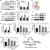
Figure 1. Post-developmental deletion of FoxO1 induces adipose browning without blocking mitochondrial turnover in mice fed on regular chow diet
(A,B) Glucose tolerance test and calculation of area under the curve (AUC) for control (Ctrl) and adO1KO mice; n=6–8. (C) Measurement of homeostasis model index of insulin resistance (HOMA-IR); n=8–12. (D–E) Analyses of UCP1 proteins in inguinal adipose tissues by immunohistochemistry (panel D, magnification, 200×) and Western blotting followed by densitometric analyses (E, n=6). (F) Measurement of body temperatures; n=6–8. (G) Transmission electron microscopic view of inguinal adipose tissue sections. The arrows point to mitochondria engulfed by autophagosome for clearance, and the asterisks mark the mitochondria not engulfed by autophagosome. (H) Counts of mitochondria inside autophagosome (MiA, marked by arrows) and mitochondria outside autophagosome (MoA, marked by asterisks) under the transmission electron microscope (area of 35 μm2); n=6–8. (I,J) Western blotting (I) and densitometric (J) analyses of FoxO1 and autophagy proteins; n=6. (K) Schematic view of how autophagic activity was sustained in adO1KO mice; *P<0.05, **P<0.01, ***P<0.001; n.s., not significant.
Figure 2
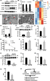
Figure 2. Post-developmental deletion of adipocyte FoxO1 activates alternate mitophagy pathways and mitochondrial biogenesis in mice fed on regular chow diet
(A,B) Western blotting (A) and densitometric (B) analyses of mitophagy adaptor and receptor proteins; n=6. (C) Illustration of the partnership between Fundc1 with Drp1 and Opa1 in connecting mitochondria to and engulfed by autophagosome via LC3II. (D,E) Western blotting (D) and densitometric (E) analyses of mitochondrial dynamics proteins Drp1, OPA1, and Mfn1; n=6. (F,G) Western blotting (F) and densitometric (G) analyses of markers of mitochondrial biogenesis; n=6. (H–J) Measurement of basal respiration (BR, H), maximal respiration (MR, I), and spare respiration capacity (SRC, J) of 3T3L1 adipocytes; n=3–4. DMSO stands for the vehicle, AS for FoxO1 inhibitor AS1842856, F1KD for Fundc1 knockdown, and P1KD for Pgc1α knockdown. (K) Schematic view of how silencing FoxO1 activates mitochondrial biogenesis and sustains mitochondrial turnover to enhance mitochondrial function. *P<0.05, **P<0.01, ***P<0.001; n.s., not significant.
Figure 3
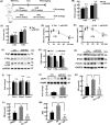
Figure 3. Post-developmental ablation of autophagy via Atg7 reduces mitochondrial turnover and causes metabolic disorders in mice fed on regular chow diet
(A,B) Western blotting (A) and densitometric (B) analyses of autophagy proteins in control (Ctrl) and adipocyte Atg7 knockout (ad7KO) mice; n=6. (C) Transmission electron microscopic view of inguinal adipose tissue sections. The arrows point to mitochondria engulfed by autophagosome for clearance, and the asterisks mark the mitochondria not engulfed by autophagosome. (D) Counts of mitochondria inside autophagosome (MiA, marked by arrows) and mitochondria outside autophagosome (MoA, marked by asterisks) under the transmission electron microscope (area of 35 μm2); n=6–8. (E) Western blotting (upper panel) and densitometric (lower panel) analyses of adipose browning maker UCP1 in inguinal adipose tissues; n=6. (F) RNAseq analysis of Atg7, mitochondrial genes (mt-Nd1, mt-Nd2, mt-Nd3, mt-Nd4, mt-cytb), fatty acid metabolism related genes (Fasn, Acaca, and Acsm3), browning marker (Cidea), and iron status marker (Fth1) in Ctrl and ad7KO mice. (G) Measurement of body temperatures; n=6. (H,I) Glucose tolerance test (H) and calculation of area under the curve (AUC, I); n=6–8. (J) Measurement of HOMA-IR; n=6–10. (K) Knockdown of Atg7 (Atg7-KD) in 3T3L1 cells; n=4–6. (L–N) Measurement of basal respiration (BR, L), maximal respiration (MR, M), and spare respiration capacity (SRC, N) of 3T3L1 adipocytes; n=3–4. (O) Schematic view of how blockage of autophagy via Atg7 impaired mitochondrial turnover and metabolic function; QC, quality control. *P<0.05, **P<0.01, ***P<0.001.
Figure 4
Figure 4. HFD disrupts autophagy and overrides the effects of adO1KO-induced adipose browning
(A) The strategies of dietary and tamoxifen treatments; CTH stands for chow→ tamoxifen→HFD, and HTH stands for HFD→ tamoxifen→HFD. (B) Measurements of body weight after the control and adO1KO mice were treated by CTH and HTH; n=8–12. (C) Measurements of fat mass after the control and adO1KO mice were treated by CTH and HTH; n=8–12. (D,E) Glucose tolerance test for the control and adO1KO mice treated by CTH (D) and HTH (E); n=6. (F,G) Western blotting (F) and densitometric (G) analyses of autophagy proteins in inguinal adipose tissues from CTH-treated mice; n=6. (H,I) Western blotting (H) and densitometric (I) analyses of mitophagy adaptor (Pink1) and receptors (Bnip3 and Fundc1) in inguinal adipose tissues from CTH-treated mice; n=6. (J) Western blotting (upper panel) and densitometric (lower panel) analyses of adipose browning maker UCP1 in inguinal adipose tissues from CTH-treated mice; n=6. (K–M) Measurement of basal respiration (BR, K), maximal respiration (MR, L), and spare respiration capacity (SRC, M) of 3T3L1 adipocytes treated with vehicle DMSO, FoxO1 inhibitor AS, and autophagy inhibitor Baf (i.e., bafilomycin A1); n=3–4; *P<0.05, **P<0.01, ***P<0.001; n.s., not significant.
Figure 5
Figure 5. Proposed mechanism by which loss of FoxO1 activates alternate mechanism to maintain mitochondrial turnover and quality control
Post-developmental deletion of FoxO1 unexpectedly up-regulates Atg7 and CTSL, concurrent with elevated Fundc1 (mitophagy receptor) and Pgc1α (inducer of mitochondrial biogenesis), which sustains mitochondrial turnover and promotes mitochondrial function and metabolic health. Blockage of Atg7 (by KD or KO), CTSL (by HFD), or autophagosome–lysosome fusion (by Bafilomycin A1, shortened as Baf), compromises mitochondrial function and metabolic health.

References

    1. Sakers A., De Siqueira M.K., Seale P. and Villanueva C.J. (2022) Adipose-tissue plasticity in health and disease. Cell 185, 419–446 10.1016/j.cell.2021.12.016 - DOI - PMC - PubMed
    1. Shi L., Tao Z., Zheng L., Yang J., Hu X., Scott K.et al. . (2023) FoxO1 regulates adipose transdifferentiation and iron influx by mediating Tgfbeta1 signaling pathway. Redox Biol. 63, 102727 10.1016/j.redox.2023.102727 - DOI - PMC - PubMed
    1. Sidossis L.S., Porter C., Saraf M.K., Borsheim E., Radhakrishnan R.S., Chao T.et al. . (2015) Browning of subcutaneous white adipose tissue in humans after severe adrenergic stress. Cell Metab. 22, 219–227 10.1016/j.cmet.2015.06.022 - DOI - PMC - PubMed
    1. Qiu Y., Sun L.Z., Hu X.L., Zhao X., Shi H.Y., Liu Z.et al. . (2020) Compromised browning plasticity of primary subcutaneous adipocytes derived from overweight Chinese adults. Diabetol. Metabolic Syndrome 12,91 10.1186/s13098-020-00599-z - DOI - PMC - PubMed
    1. Carey A.L., Formosa M.F., Van Every B., Bertovic D., Eikelis N., Lambert G.W.et al. . (2013) Ephedrine activates brown adipose tissue in lean but not obese humans. Diabetologia 56, 147–155 10.1007/s00125-012-2748-1 - DOI - PubMed

Publication types

MeSH terms

Substances