. 2024 Jun;26(6):101120.
doi: 10.1016/j.gim.2024.101120. Epub 2024 Mar 10.

Biallelic USP14 variants cause a syndromic neurodevelopmental disorder

Affiliations

Biallelic USP14 variants cause a syndromic neurodevelopmental disorder

Frédéric Ebstein et al. Genet Med. 2024 Jun.

Abstract

Purpose: Imbalances in protein homeostasis affect human brain development, with the ubiquitin-proteasome system (UPS) and autophagy playing crucial roles in neurodevelopmental disorders (NDD). This study explores the impact of biallelic USP14 variants on neurodevelopment, focusing on its role as a key hub connecting UPS and autophagy.

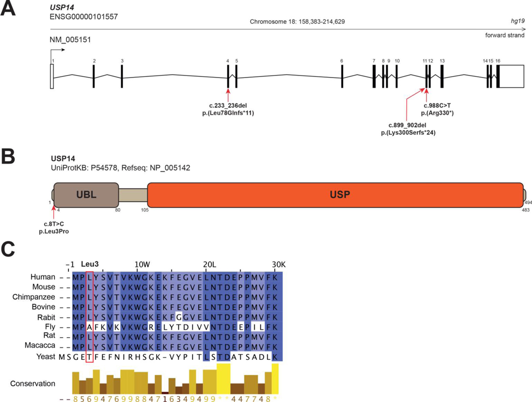
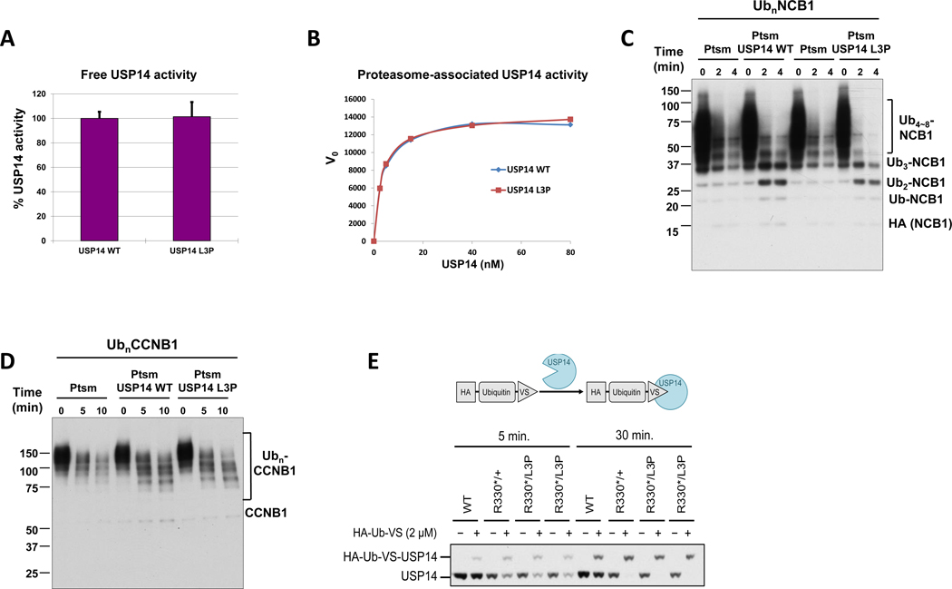
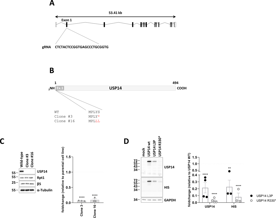
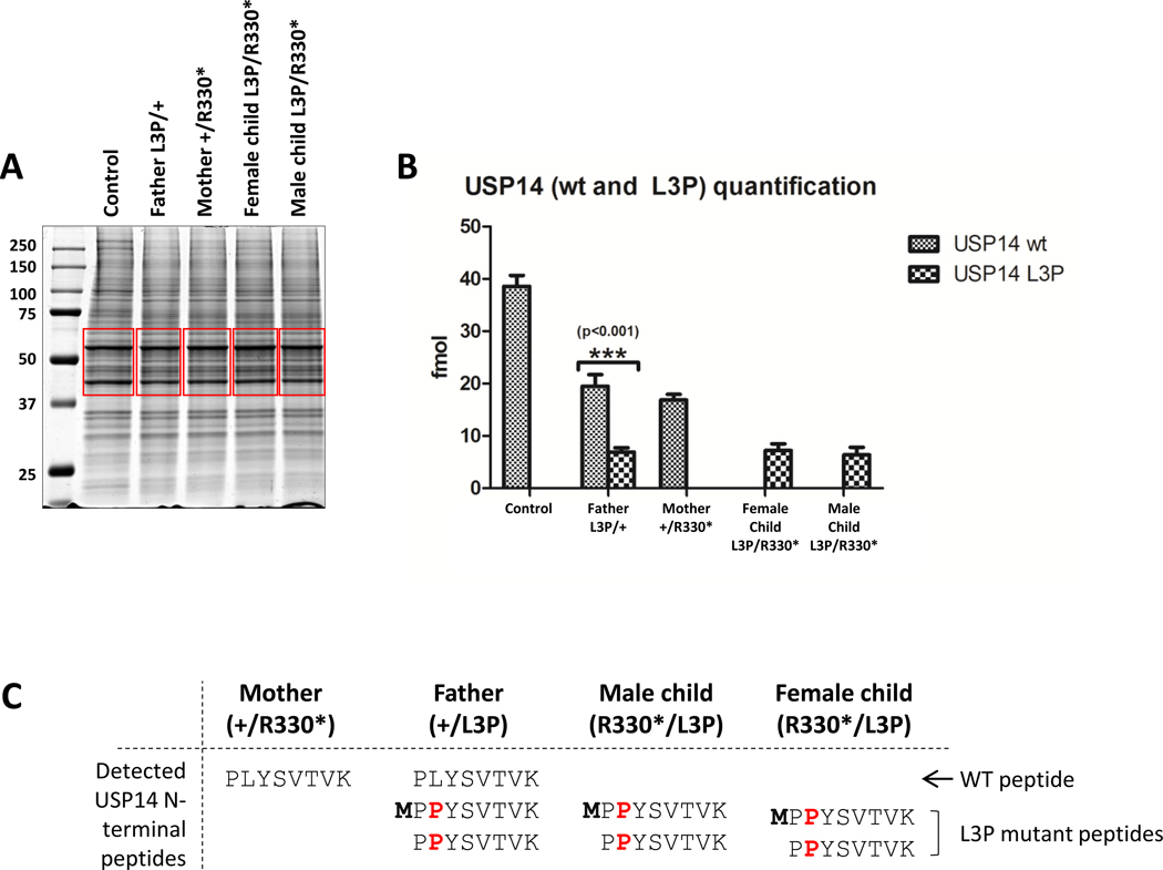
Methods: Here, we identified biallelic USP14 variants in 4 individuals from 3 unrelated families: 1 fetus, a newborn with a syndromic NDD and 2 siblings affected by a progressive neurological disease. Specifically, the 2 siblings from the latter family carried 2 compound heterozygous variants c.8T>C p.(Leu3Pro) and c.988C>T p.(Arg330∗), whereas the fetus had a homozygous frameshift c.899_902del p.(Lys300Serfs∗24) variant, and the newborn patient harbored a homozygous frameshift c.233_236del p.(Leu78Glnfs∗11) variant. Functional studies were conducted using sodium dodecyl-sulfate polyacrylamide gel electrophoresis, western blotting, and mass spectrometry analyses in both patient-derived and CRISPR-Cas9-generated cells.

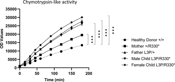
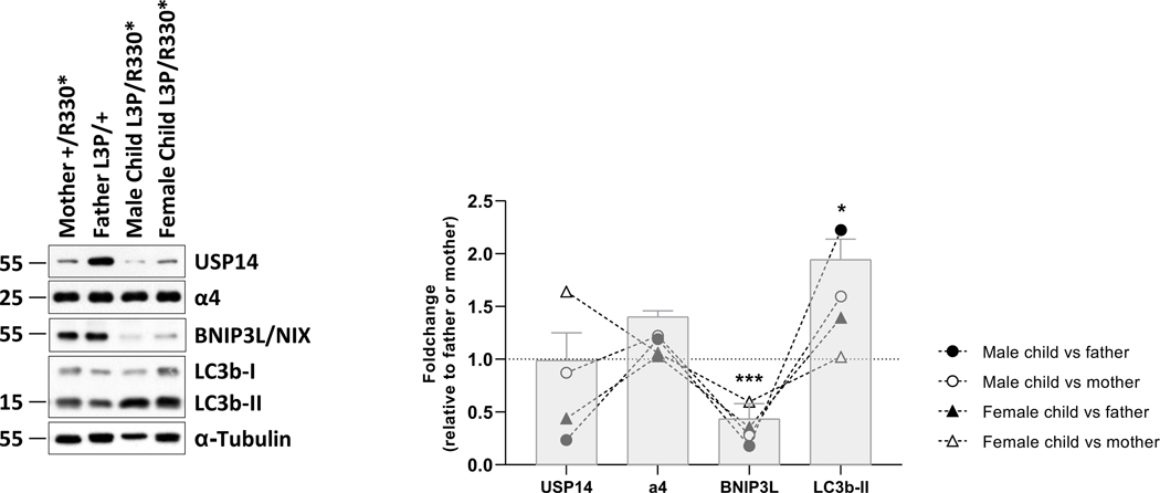
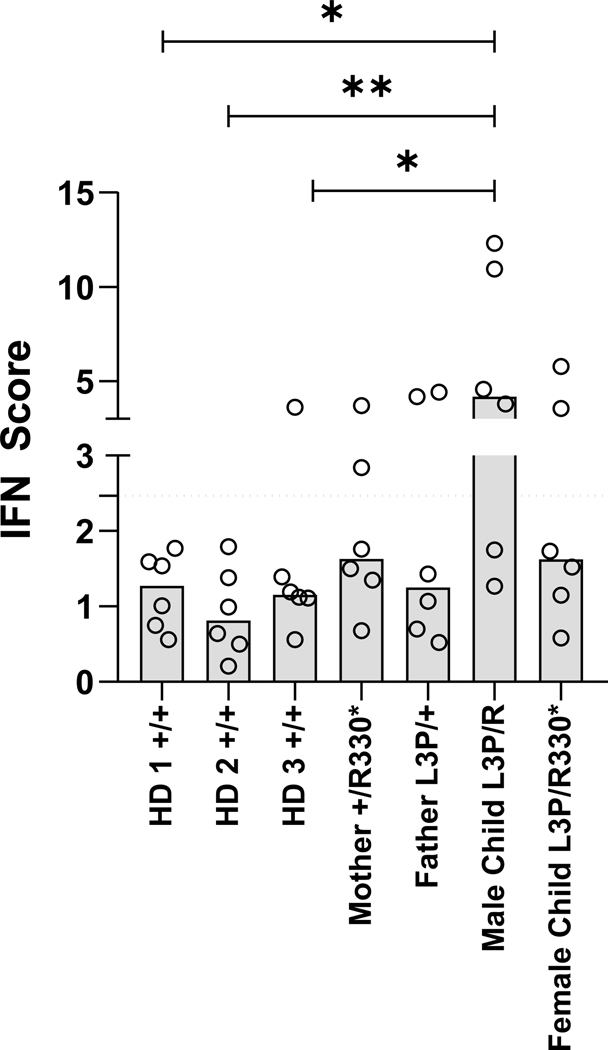
Results: Our investigations indicated that the USP14 variants correlated with reduced N-terminal methionine excision, along with profound alterations in proteasome, autophagy, and mitophagy activities.

Conclusion: Biallelic USP14 variants in NDD patients perturbed protein degradation pathways, potentially contributing to disorder etiology. Altered UPS, autophagy, and mitophagy activities underscore the intricate interplay, elucidating their significance in maintaining proper protein homeostasis during brain development.

Keywords: Loss-of-function variants; N-terminal methionine excision; Neurodevelopmental disorders; USP14; Ubiquitin-proteasome system.


Conflict of interest statement

Conflict of Interest The authors declare no conflicts of interest.

Figures

Figxure 1.
Figxure 1.. USP14 variants.
A. Exon structure of longest USP14 transcript, containing 16 exons (NM_005151). Nonsense and frameshift variants identified in the present study are indicated with red arrows. B. Protein structure of USP14 (UniProtKB - P54578). Functional domains (PROSITE annotation rule, UniProtKB/Swiss-Prot format) are indicated. UBL: Ubiquitin-like domain. USP: Ubiquitin specific protease domain. Nonsynonymous variant c.8T>C, (p.Leu3Pro); (individuals 1 and 2) is indicated with a red arrow. C. Multiple sequence alignment of USP14 orthologs from 8 eukaryotic species compared to USP14 human protein sequence. Clustal W multiple sequence alignment program. Conservation scores are mentioned below the alignment. Red boxes identify variant positions.
Figure 2.
Figure 2.. Subjects with UPS14 variants exhibit increased proteasome activity.
Ten micrograms of native cell lysates generated from T cells of a healthy donor (HD) and subjects carrying the p.(Arg330*) and/or p.(Leu3Pro) USP14 variants were incubated at 37°C on a 96-well plate in a final 100 μl volume containing 0.1 mM of the Suc-LLVY-AMC fluorogenic peptide. Fluorescence activity reflecting proteasome chymotrypsin-like activity was measured at 360/460 nm on a microplate reader over a 180-min period of time, as indicated. ***p<0.001 (unpaired two-tailed t test, n=4).
Figure 3.
Figure 3.. Deubiquitinating activity of recombinant USP14-L3P.
A. Ub-AMC hydrolysis assays with free forms of USP14 WT and L3P at 1.5 μM. Error bars represent S.D. from triplicate experiments. B. Kinetic analysis of proteasome-bound USP14 WT and L3P activities with Ub-AMC as substrate. Graded concentrations of recombinant USP14 were reconstituted with 1 nM Ub-VS-treated proteasome, and cleavage of Ub-AMC (1 μM) by USP14 was measured over 80 min in real time. The plots shown represent mean values of initial reaction rates from triplicate experiments. C. In vitro deubiquitination/degradation assays with UbnNCB1, human proteasome (4 nM), and WT or L3P USP14 (80 nM). D. In vitro deubiquitination/degradation assays with UbnCCNB1, human proteasome (7 nM), and WT or L3P USP14 (140 nM). E. The irreversible covalent modifier ubiquitin vinyl sulfone, carrying an HA epitope tag, was added at 2 micromolar to lysates from patient fibroblasts, and the samples incubated at 25°C for either 5 or 30 min as indicated. Samples were then analyzed by SDS-PAGE and immunoblotting, using an antibody to USP14. Each lane was loaded with 25 micrograms of lysate. HA-Ub-VS reacts with the catalytic cysteine of proteasome-activated USP14.
Figure 4.
Figure 4.. Patients with UPS14 biallelic variants are associated with alterations of the autophagy/mitophagy pathways.
Left panel: ten to twenty micrograms of RIPA cell lysates obtained from T cells of subjects with USP14 variants were separated by SDS-PAGE prior to western-blotting using antibodies specific for USP14, α4, BNIP3L/NIX, LC3B and α-tubulin (loading control), as indicated. Shown are representative western-blots of three independent experiments performed. Right panel: Shown is the quantification of the Western-blots by densitometry. Data are presented as foldchanges in the affected male and female children vs their father and/or mother whose densitometric measurements were set to 1 (gridline), as indicated. Columns indicate the foldchange mean values ± SEM calculated from the normalizations of three independent biological replicates. Statistical significance was assessed by unpaired Student’s test (*p<0.05 and ***p<0.001).
Figure 5.
Figure 5.. The p.(Arg330*) nonsense mutation fails to give rise to a USP14 truncated variant in USP14−/− SH-SY5Y cells.
A. Schematic representation of the USP14 gene as well as the region in exon 1 targeted by the specific guide RNA used for gene deletion by CRISPR/Cas9. B. Primary structures generated from the sequencing results of USP14 PCR amplicons obtained for wild-type SHSY-5Y cells and clones #3 and #16, as indicated. C. Left panel: wild-type SHSY-5Y cells and clones #3 and #16 were subjected to SDS-PAGE and western-blotting using antibodies specific for USP14, Rpt1 and Beta5, as indicated. Equal protein loading was ensured by using an antibody directed to α-tubulin. Right panel: Shown is the quantification of the Western-blots by densitometry. Data are presented as foldchanges (n=3) in clones #3 and #16 vs the SHSY-5Y parental cell line whose densitometric measurements were set to 1 (gridline), as indicated. Statistical significance was assessed by unpaired Student’s test (****p<0.0001). D. Left panel: SHSY-5Y clone #16 was subjected to a 24-h transfection with expression vectors coding for HIS-tagged wild-type UPS14 as well as the p.(Arg330*) or p.(Leu3Pro) USP14 variants. Overexpressed proteins were quantified by SDS-PAGE/western-blotting using anti-USP14, anti-HIS and anti-GAPDH (loading control) antibodies, as indicated. Right panel: Shown is the quantification of the Western-blots by densitometry. Data are presented as foldchanges of the USP14 and HIS signals in SHSY-5Y cells transected with the p.(Leu3Pro) (black circles) and p;(Arg330*) (white circles) USP14 variants relative to SHSY-5Y cells transected with wild-type USP14 whose densitometric measurements were set to 1 (gridline), as indicated. Statistical significance was assessed by unpaired Student’s test (**p<0.01 and ****p<0.0001).
Figure 6.
Figure 6.. Mass spectral analysis of whole-cell lysates derived from patient cells reveals decreased expression and N-terminal methionine processing of the Leu3Pro (L3P) USP14 variant.
A. Coomassie-stained SDS-PAGE of protein lysates from subjects carrying the p.(Arg330*) and/or p.(Leu3Pro) USP14 variants, as indicated. Gel slices excised for trypsin digestion and subsequent mass spectrometry analysis are indicated in red. B. Quantification of wild-type (wt) and Leu3Pro (L3P) USP14 peptides by MS using a PRM (parallel reaction monitoring) method. C. Table recapitulating the identification of N-terminal methionine untrimmed and trimmed USP14 wild-type (WT)- and Leu3Pro-derived peptides found in USP14 immuno-precipitates of each sample, as indicated.
Figure 7.
Figure 7.. UPS14 loss-of-function is not consistently associated with sterile type I IFN responses.
Interferon (IFN) scores of whole-blood samples collected and stabilized in PAXgene RNA tubes from three unrelated healthy individuals (HD) and subjects carrying the p.(Arg330*) and/or p.(Leu3Pro) USP14 variants, as indicated. Scores were calculated as the median of the relative quantification (RQ) of seven ISG (i.e., IFIT1, IFI27, IFI44L, ISG15, RSAD2 and SIGLEC1) over a single calibrator control, as measured by RT-qPCR (see Materials & Methods). Statistical significance was assessed by paired ratio t test (*p<0.05 and **p<0.01).

References

    1. Rock KL, Gramm C, Rothstein L, et al. Inhibitors of the proteasome block the degradation of most cell proteins and the generation of peptides presented on MHC class I molecules. Cell. 1994;78(5):761–771. doi:10.1016/S0092-8674(94)90462-6 - DOI - PubMed
    1. Wilkinson KD. Regulation of ubiquitin-dependent processes by deubiquitinating enzymes. FASEB J. 1997;11(14):1245–1256. doi:10.1096/FASEBJ.11.14.9409543 - DOI - PubMed
    1. Borodovsky A, Kessler BM, Casagrande R, Overkleeft HS, Wilkinson KD, Ploegh HL. A novel active site-directed probe specific for deubiquitylating enzymes reveals proteasome association of USP14. EMBO J. 2001;20(18):5187–5196. doi:10.1093/EMBOJ/20.18.5187 - DOI - PMC - PubMed
    1. Li T, Naqvi NI, Yang H, Teo TS. Identification of a 26S proteasome-associated UCH in fission yeast. Biochem Biophys Res Commun. 2000;272(1):270–275. doi:10.1006/BBRC.2000.2767 - DOI - PubMed
    1. Yao T, Cohen RE. A cryptic protease couples deubiquitination and degradation by the proteasome. Nature. 2002;419(6905):403–407. doi:10.1038/NATURE01071 - DOI - PubMed

Publication types

Substances