. 2024 Jun 10;45(6):436-449.
doi: 10.1093/carcin/bgae022.

Growth modulatory effects of fenretinide encompass keratinocyte terminal differentiation: a favorable outcome for oral squamous cell carcinoma chemoprevention

Affiliations

Growth modulatory effects of fenretinide encompass keratinocyte terminal differentiation: a favorable outcome for oral squamous cell carcinoma chemoprevention

Daren Wang et al. Carcinogenesis. .

Abstract

Oral squamous cell carcinoma (OSCC) is worldwide health problem associated with high morbidity and mortality. From both the patient and socioeconomic perspectives, prevention of progression of premalignant oral intraepithelial neoplasia (OIN) to OSCC is clearly the preferable outcome. Optimal OSCC chemopreventives possess a variety of attributes including high tolerability, bioavailability, efficacy and preservation of an intact surface epithelium. Terminal differentiation, which directs oral keratinocytes leave the proliferative pool to form protective cornified envelopes, preserves the protective epithelial barrier while concurrently eliminating growth-aberrant keratinocytes. This study employed human premalignant oral keratinocytes and an OSCC cell line to evaluate the differentiation-inducing capacity of the synthetic retinoid, fenretinide (4HPR). Full-thickness oral mucosal explants were evaluated for proof of concept differentiation studies. Results of this study characterize the ability of 4HPR to fulfill all requisite components for keratinocyte differentiation, i.e. nuclear import via binding to cellular RA binding protein-II (molecular modeling), binding to and subsequent activation of retinoic acid nuclear receptors (receptor activation assays), increased expression and translation of genes associated with keratinocyte differentiation [Reverse transcription polymerase chain reaction (RT-PCR), immunoblotting] upregulation of a transglutaminase enzyme essential for cornified envelope formation (transglutaminase 3, functional assay) and augmentation of terminal differentiation in human oral epithelial explants (image-analyses quantified corneocyte desquamation). These data build upon the chemoprevention repertoire of 4HPR that includes function as a small molecule kinase inhibitor and inhibition of essential mechanisms necessary for basement membrane invasion. An upcoming clinical trial, which will assess whether a 4HPR-releasing mucoadhesive patch induces histologic, clinical and molecular regression in OIN lesions, will provide essential clinical insights.


Figures

Graphical Abstract
Graphical Abstract
Figure 1.
Figure 1.
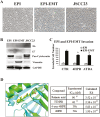
Characterization of short tandem repeat-authenticated EPI, EPI-EMT and JSCC23 cell lines. (A) Phase contrast images of log growth cultures 100× image (Nikon DS-Ri1 color digital microscope camera). While all cell lines exhibited high proliferation indices in culture, qualitative differences were noted. The EPI cell line exhibits a uniform nuclear appearance and a relatively higher amount of cytoplasm. In contrast, the EPI-EMT cultures show a more spindly, angular phenotype. Finally, the JSCC23 cell line, which was derived from a primary OSCC with lymph node metastases, demonstrates higher nuclear-to-cytoplasmic ratios, greater nuclear and cellular pleomorphism and loss of contact inhibition. (B) Immunoblotting was used to characterize selective intermediate filament proteins and presence or absence of oncogenic HPV E6 and E7 proteins. Consistent with their epithelial origin, all cell lines expressed pancytokeratin and also showed concurrent vimentin expression. Vimentin levels, however, were highest in the EPI-EMT cells which also possessed the most spindled ‘fibroblast like’ appearance (30). All cell lines expressed at least one oncogenic HPV E6 or E7 protein. The EPI-EMT cells were the exclusive line that expressed both E6 and E7. (C) Collagen type IV simulated basement membrane invasion assays were conducted to assess whether or not the EPI-EMT phenotypic appearance correlated with enhanced invasive capacity. Log growth cells received the following treatments: (i) Control 0.01% DMSO; (ii) 1 µM 4HPR; (iii) 1 µM ATRA for 6 days. To optimize cell responsiveness to chemoattractants, 24 h prior to the invasion assay, cells were cultured in BASE medium (same as above without serum) with the same treatments. Cells were then seeded onto ECMatrixTM-coated microporous polycarbonate membrane (Millipore QCM™ 96-well Invasion Assay, Massachusetts). The lower chamber contained conditioned medium JSCC3. After 18 h of invasion (37°C, 5% CO2), cell detachment buffer was used to dislodge successfully invaded cells. Cells were subsequently lysed and detected by CyQuant GR dye. Invaded cell number was determined relative to the fluorescently labeled, cell-line-specific standard curve by running a fluorescent cell standard curve. Fluorescence was assessed using a FLUOstar Omega (BMG LABTECH) fluorescence plate reader using 480/520 nm filter set. Assay results confirmed a markedly enhanced invasiveness in the EPI-EMT cell lines and also confirmed the capacity of both 4HPR and ATRA to significantly inhibit invasion (mean + SEM, EPI n = 4, EPI-EMT n = 5, * P < 0.05, Kruskal–Wallis with Dunn’s Multiple Comparisons post hoc test). (D) Molecular modeling studies confirm the capacity for 4HPR to undergo nuclear translocation. In order for retinoids to impact gene expression, transportation to the nucleus is essential. RA and its derivatives employ binding to the cellular RA binding protein, CRABP-II, for successful nuclear importation. Our molecular modeling data confirm that oxidized 4HPR can bind to and function as competitive inhibitors of RA binding the CRABP-II. Superimposition of 4HPR (background, yellow) and 1CBS (foreground, green). The 4HPR structure extends deeper into the protein's binding pocket due to the additional length from the 4-hydroxyphenyl group where if OH is placed in the same location of the CO2 of the RA.
Figure 2.
Figure 2.
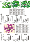
4HPR activates the nuclear transcription regulatory RARs. The capacity for 4HPR to bind (molecular modeling assessment) and activate the RARs was assessed using RA reporter assay system. This system employs human cells that express high levels of RARs and possess a luciferase reporter gene. Quantification of luciferase expression, therefore, provides a surrogate measure of RAR activity in treated cells. (A.i) Molecular modeling studies showed that in all cases, the docking with the endogenous ligand matched the pose from the crystal structure. 4HPR and 4-oxo-4HPR, bind with similar affinities as RA or 9-cis-RA indicating that these compounds will be a competitive inhibitor for RAR, particularly the RARa isoform. Model compounds labeled as 9-cis-RA (green), RA (red), 4-oxo-4HPR (blue) and 4HPR (cyan). (A.ii) Following a 24 h incubation the reporter cells were treated with 1 µM of 4HPR, 4-oxo-HPR, ATRA and 9-cis-RA. Adapalene served as the positive assay control. All agents except 4-oxo-HPR significantly increased RAR receptor activity in all three isoforms, α, β and γ. Mean + SEM, n = 8, *P < 0.05, **P < 0.010. Kruskal–Wallis, Dunns multiple comparison post hoc test. (B.i) Molecular modeling studies to assess 4HPR and it oxidized metabolite 4-oxo-HPR revealed that 4HPR (green) does not bind in the same pocket as the others and has a significantly smaller Kd. Model compounds labeled as 4HPR (green) in the binding pocket with the endogenous ligand 9-cis-RA (pink) and 9-cis-4HPR (Yellow). (B.ii) Cells were handled in an identical fashion as that described for the RAR studies. The ATRA and 9-cis-RA provided strong, positive induction of all three isoforms (RARa, RARb and RARg) of the RXR receptors. In contrast, neither 4HPR nor 4-oxo-HPR demonstrated any inductive effect. Mean + SEM, n = 8, **P < 0.01 Kruskal–Wallis, Dunns multiple comparison post hoc test.
Figure 3.
Figure 3.
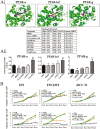
4HPR does not activate the PPARs nor induce cell proliferation. The capacity of 4HPR to bind (molecular modeling assessment) and activate PPARs was assessed using the PPAR reporter assay system. (A.i) Molecular modeling compounds are labeled as 4HPR (blue) and 4-oxo-HPR (red) in the binding pocket with the endogenous ligand 9-cis-RA (yellow). 4HPR does not bind in the same pocket as the others and has a significantly smaller Kd. Modeling studies indicated that 4HPR interacted with the PPAR binding site on all isoforms, comparable to moderate to high potency PPAR targeted inhibitors, e.g. GW2331. (A.ii) CHO cells were treated with 1 µm 4HPR, 4-oxo-HPR, ATRA and 9-cis-RA for 24 h, followed by assessment of PPAR activation using Indigo reporter assay kit. Despite comparable calculated molecular binding affinities, 9-cis-RA and 4HPR exhibited different effects on PPAR isoform signaling. 9-cis-RA significantly increased receptor activation (P < 0.01) in all three isoforms PPARa, PPARb/d and PPARg whereas 4HPR significantly repressed signaling for the PPARa and PPARg isoforms, (#P < 0.05, respectively). Addition of ATRA also significantly increased the g isoform signaling (P < 0.01). Mean + SEM, n = 8, Kruskal–Wallis with a Dunns multiple comparison post hoc test. (B) The effects of RA derivatives on cell proliferation were determined over a 6-day time course. Cells were treated with 4HPR, 4-oxo-HPR, ATRA and 9-cis-RA at doses of 1 and 2.5 µM, with fresh medium and RA derivatives provided every other day. Cell numbers were determined using BioRad TC20 Automated Cell Counter (BioRad, Hercules, CA). The cell growth curves demonstrated that the EPI-EMT cells displayed the highest growth rate. In none of the cultures, at any retinoid dose, did the addition of retinoids augment cells proliferation. The established apoptosis inducer, 4-oxo-HPR significantly decreased cell numbers in the EPI and EPI-EMT cells at 1 µM. All three cell lines showed significant growth inhibition at day 6 with the addition of 1.0 µM 4-oxo-HPR (P < 0.05) and 2.5 µM 4-oxo-HPR (**P < 0.01) n = 3, Kruskal–Wallis with a Dunns multiple comparison post hoc test.
Figure 4.
Figure 4.
4HPR modulates gene expression and protein translation toward keratinocyte differentiation. (A) Addition of low dose 4HPR (1 µM, 48 h treatment) induced a statistically significant increase in genes associated with terminal differentiation, retinoic metabolism, retinoid signaling and retinoid transport. One of the 4HPR upregulated genes (CYP26A1) is known to be regulated by RA. The fold change in expression was determined in accordance the delta CT calculation (n = 3) (31). (B) Time course studies to assess the impact of low dose 4HPR (1 µM) on cell production of proteins associated with retinoid-associated nuclear translocation, gene expression, metabolism and terminal differentiation were conducted. 4HPR-administrated periods were specified to 1, 2, 4 and 6 days, respectively, to each cell lines for expression assessment; same concentration (volume ratio) of DMSO treatment were served as control (see Supplementary Table 2, available at Carcinogenesis online).
Figure 5.
Figure 5.
4HPR augments functional activity of a key differentiation enzyme, transglutaminase 3. Studies were conducted to assess the effects of low dose 4HPR (1 µM, 96 h, fresh medium and 4HPR every other day) on the enzymatic function of two transglutaminases [TGM1 (A) and TGM3 (B)] that are essential for cornified envelope formation and keratinocyte terminal differentiation. All enzymatic functional assays (Zedira transglutaminase fluorogenic assay, Darmstadt, Germany) were conducted concurrently, using the same 96-well black polystyrene microplate. Our data confirm that both the EPI and EPI-EMT cell lines exhibited basal levels of both TGM1 and TGM3 activity, with greater activity demonstrated by the EPI cells. Treatment with 4HPR resulted in a significant increase in TGM3 function in both the EPI and EPI-EMT cultures. *P < 0.05, n = 3, two-tailed, non-paired Wilcoxon rank-sum test. While the JSCC23 cells also exhibited basal level TGM1 and TGM3 activity, no upregulation was noted with 4HPR treatment (data not shown).
Figure 6.
Figure 6.
4HPR promotes corneocyte formation and desquamation in oral mucosal explants. (A) MatTek EpiOral explant treatment with 1 µM 4HPR increased keratinocyte differentiation as evidenced by formation of terminally differentiated keratinocytes undergoing superficial desquamation. Although control explants displayed some keratinocyte desquamation, the extent in the control tissues was negligible relative to 4HPR-treated explants. Notably, keratin envelope formation and corneocyte desquamation was observed in the 4HPR-treated explants regardless of fixation method (OCT or formalin–paraffin embedded) employed. In addition, stratified squamous intact epithelium remained underlying the desquamated keratinocytes. Microscopic image analyses to quantify desquamated keratinocytes confirmed 4HPR significantly increased keratinocyte terminal differentiation and desquamation explants. (mean ± SD; **P< 0.01; 4HPR bolus delivery, n = 8; no 4HPR control, n = 3; Kruskal–Wallis, Dunns multiple comparison post hoc test). (B) Diffuse cytosolic staining for TGM1 is present in both the 4HPR treated and control explants. Immunohistochemical stains for the other two cytosolic proteins were comparable regardless of 4HPR treatment. (C) Ki-67 nuclear staining (highlighted by arrows), indicative of cell proliferation/explant viability, is observed in some of the non-desquamated basal layer keratinocytes in both the control and 4HPR treated explants.

Similar articles

References

    1. Coletta, R.D. et al.. (2020) Grand challenges in oral cancers. Front. Oral Health, 1, 3. - PMC - PubMed
    1. Suresh, G.M. et al.. (2019) Prognostic indicators of oral squamous cell carcinoma. Ann. Maxillofac. Surg., 9, 364–370. - PMC - PubMed
    1. Ribeiro-Rotta, R.F. et al.. (2022) The cost of oral cancer: a systematic review. PLoS One, 17, e0266346. - PMC - PubMed
    1. Sporn, M.B. et al.. (1976) Approaches to prevention of epithelial cancer during the preneoplastic period. Cancer Res., 36, 2689–2702. - PubMed
    1. Sporn, M.B. et al.. (2002) Chemoprevention: an essential approach to controlling cancer. Nat. Rev. Cancer, 2, 537–543. - PubMed

MeSH terms