. 2024 Apr 9;92(4):e0003724.
doi: 10.1128/iai.00037-24. Epub 2024 Mar 12.

Corrected and republished from: "Extracellular Vesicle Formation in Cryptococcus deuterogattii Impacts Fungal Virulence"

Affiliations

Corrected and republished from: "Extracellular Vesicle Formation in Cryptococcus deuterogattii Impacts Fungal Virulence"

Rafael F Castelli et al. Infect Immun. .

Abstract

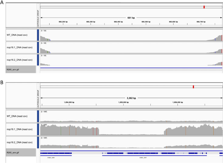
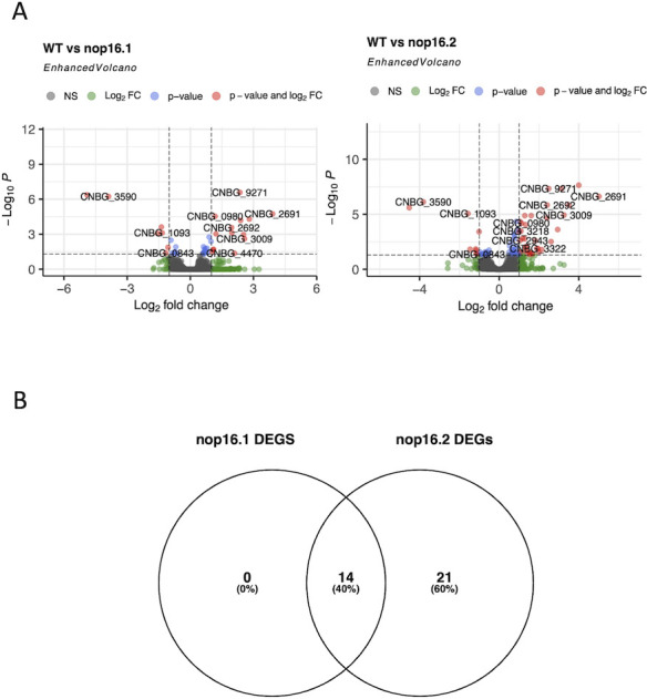
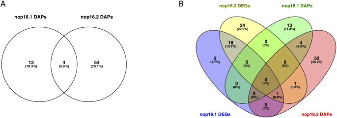
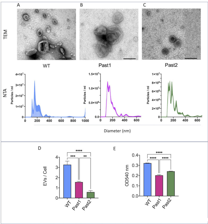
Small molecules are components of fungal extracellular vesicles (EVs), but their biological roles are only superficially known. NOP16 is a eukaryotic gene that is required for the activity of benzimidazoles against Cryptococcus deuterogattii. In this study, during the phenotypic characterization of C. deuterogattii mutants expected to lack NOP16 expression, we observed a reduced EV production. Whole-genome sequencing, RNA-Seq, and cellular proteomics revealed that, contrary to our initial findings, these mutants expressed Nop16 but exhibited altered expression of 14 genes potentially involved in sugar transport. Based on this observation, we designated these mutant strains as Past1 and Past2, representing potentially altered sugar transport. Analysis of the small molecule composition of EVs produced by wild-type cells and the Past1 and Past2 mutant strains revealed not only a reduced number of EVs but also an altered small molecule composition. In a Galleria mellonella model of infection, the Past1 and Past2 mutant strains were hypovirulent. The hypovirulent phenotype was reverted when EVs produced by wild-type cells, but not mutant EVs, were co-injected with the mutant cells in G. mellonella. These results connect EV biogenesis, cargo, and cryptococcal virulence.

Keywords: Cryptococcus; extracellular vesicles.


Conflict of interest statement

The authors declare no conflict of interest.

Figures

Fig 1
Fig 1
Tentative generation of null mutants of NOP16 in C. deuterogattii. (A) NOP16 inactivation strategy. TV is the representation of the targeting vector constructed by the Delsgate methodology, with 5 NOP16 and 3 NOP16 representing the 5′ and 3′ gene flanks of the NOP16 gene, respectively. The primers used to amplify the 5′ (5F and 5R) and 3′ (3F and 3R) of NOP16 are represented as arrowheads. NATR is the cassette that confers nourseothricin resistance. (B) Nourseothricin-resistant cells were evaluated for the presence of the NOP16 gene using internal diagnostic primers (IF and IR), using the ACT gene as a loading control. (C) WT and two null mutants were evaluated for their sensitivity to mebendazole. Bars represent the average of the ratio between growth in 1 mM mebendazole normalized to the growth in a drug-free medium obtained in three independent experiments. Mutant cells display decreased sensitivity to mebendazole (**P < 0.01 and ***P < 0.0001) as revealed by analysis of variance followed by Dunnett’s multiple-comparison analysis. In contrast to what these results suggested, NOP16 deletion was not achieved. Please see Results and Fig. 2 to 8 for a detailed explanation and further correction.
Fig 2
Fig 2
Transcriptional profile (A) and genomic structure (B) of the NOP16 (CNBG_3695) gene in the analyzed strains. (A) IGV panel illustrating the coverage of aligned RNA-Seq reads (read cov) of the WT (upper), nop16.1 (middle), and nop16.2 (bottom) strains. The blue arrows below the histogram panel represent the gene architecture. (B) Genomic structure of the NOP16 (CNBG_3695) locus in the analyzed strains. IGV panel illustrating the coverage of aligned DNA Seq reads (read cov) of the WT (upper), nop16.1 (middle), and nop16.2 (bottom) mutants. The blue arrows below the histogram panel represent the gene architecture.
Fig 3
Fig 3
Genomic structure of CNBG_3591 locus in the analyzed strains. IGV panel illustrating the DNA Seq aligned reads to the WT (upper), nop16.1 (middle), and nop16.2 (bottom) mutants. The blue arrows below the histogram panel represent the gene architecture of CNBG_3591 (center) and the partial architecture of Fig. 3 genomic structure of the CNBG_3591 locus in the analyzed strains. IGV panel illustrating the coverage of aligned DNA Seq reads (read cov) of the WT (upper), nop16.1 (middle), and nop16.2 (bottom) mutants. The blue arrows below the histogram panel represent the CNBG_3591 gene architecture as well as the partial maps of surrounding genes CNBG_3590 (left) and CNBG_3592 (right).
Fig 4
Fig 4
Predicted deleted (A), tandem-duplicated (B) loci specific to nop16.1 and nop16.2. (A) IGV panel illustrating the coverage of aligned DNA Seq reads (read cov) of the WT (upper), nop16.1 (middle), and nop16.2 (bottom) mutants. (B) IGV panel illustrating the coverage of aligned DNA Seq reads (read cov) of the WT (upper), nop16.1 (middle), and nop16.2 (bottom) mutants.
Fig 5
Fig 5
Comparative analysis of gene expression in WT and mutant strains. (A) PCA of transformed data from DESeq2, highlighting the analyzed groups. (B) Venn diagram highlighting the shared and specific DEGs from the comparison between WT and nop16.1 or nop16.2 strains.
Fig 6
Fig 6
Regulation of gene expression in the mutant strains of C. deuterogattii. (A) DEGs profile presented by nop16 mutants. Data from DESeq2 were plotted using the EnhancedVolcano (7) package to evaluate the set of DEGs found between the comparison of WT and nop16.1 mutant (left) or WT and nop16.2 mutant (right). (B) Venn diagram highlighting the positively regulated genes in WT compared to nop16.1 or nop16.2 mutants.
Fig 7
Fig 7
DAPs profiles presented by the nop16.1 mutant compared to the WT strain (A) or nop16.2 mutant compared to the WT strain (B). Data were processed using the PatternLab V pipeline and plotted after applying the TFold function. The F-stringency was set to 0.45, and the q-value cutoff value was 0.05. Blue dots represent DAPs.
Fig 8
Fig 8
Venn diagrams highlighting the shared and specific DAPs from the comparison between WT and nop16.1 or nop16.2 mutants (A) and the shared and specific DAPs and DEGs from the comparison between WT and nop16.1 or nop16.2 mutants (B).
Fig 9
Fig 9
C. deuterogattii mutant strains manifest a hypovirulent phenotype. Infection of G. mellonella with the independent Past mutants 1 (A) and 2 (B) resulted in a smaller efficacy in killing the animals, in comparison with wild-type (WT) cells (P < 0.0001 for both mutants). Control systems were injected with phosphate buffered saline (PBS) only. Statistical analysis was performed with the Mantel-Cox test.
Fig 10
Fig 10
Analysis of growth rates (A–C) and ultrastructural aspects (D–F) of wild-type (WT) and mutant cells. Independently of the use of Sabouraud (A), YPD (B), or RPMI (C) as the growth media, the mutant strains always manifested higher proliferation rates, in comparison to WT cells. Analysis of the ultrastructural features of WT (D), Past1 (E), and Past2 (F) cells revealed no evident alterations. Scale bars in D–F correspond to 500 nm.
Fig 11
Fig 11
Analysis of the surface architecture of C. deuterogattii and its impact on fungal phagocytosis. Fluorescence microscopy analysis of the cell surface of WT (A), Past1 (B), and Past2 (C) cells revealed similar aspects of cell wall chitin (blue fluorescence), chitooligomers (green fluorescence), and the capsule (red fluorescence) in the three strains. GXM secretion tended to be higher in the Past1 mutant (**P = 0.0053), but not the Past2 strain (D; ns, not significant). India ink counterstaining of WT (E), Past1 (F), and Past2 (G) cells suggested similar capsular dimensions, but the determination of the capsule sizes revealed lower average values for the mutant cells (H, ****P < 0.0001, with a 95% confidence level of 95.61%). SEM was also used for the observation of the capsules of WT (I), Past1 (J), and Past 2 (K) cells, revealing similar capsular structures. The phagocytosis rates of the three strains by mouse macrophages were also determined (L). No significant (ns) differences between the three strains were observed.
Fig 12
Fig 12
Cell wall (calcofluor white, CFW) and Golgi staining (C6-NBD-Cer) in WT and mutant cells. (A) Microscopic examination of stained fungal cells suggested reduced levels of Golgi staining in mutant cells. (B) Quantitative determination of fluorescent cells (100 cells for each condition) confirmed a significantly reduced detection of Golgi staining in the mutants, in comparison with WT cells. Scale bars, 10 µm.
Fig 13
Fig 13
Analysis of EV formation in C. deuterogattii. EVs produced by wild-type (A), Past1 (B), and Past2 (C) cells were characterized by TEM (upper panels) and NTA (lower panels). EVs from all strains, in general, manifested similar properties. (D) Quantification of EVs produced by each strain revealed a significantly reduced number of vesicles produced by mutant cells, in comparison to the WT strain (****P  <  0.0001; ***P  =  0.002). EV production was smaller in the Past2 mutant than in the Past1 strain (**P  =  0.0036). (E) Measurement of urease activity in the three strains revealed that the mutants had a lower ability to hydrolyze urea (****P  <  0.0001). Both mutants manifested similar urease activities (ns, not significant).
Fig 14
Fig 14
Multivariate data analysis of small molecule composition in WT and mutant cells of C. deuterogattii based on liquid chromatography-mass spectrometry (LC-MS) data. (A) PLS-DA of EVs LC-MS data. Each sphere in this analysis represents a single replicate. The elliptical area around the three replicates represents the confident zone of each group. In this analysis, R2 and Q2 values corresponded to 0.98 and 0.20, respectively, confirming the consistency between the original and cross-validation predicted data. (B) Three metabolites [Phe-Pro, Pyro Glu-Leu, and Cyclo (Tyr-Pro)] were classified as variables important in the projection (VIP) features indicated by PLS-DA. Only VIPs with a coefficient score above 3.0 and P < 0.01 for the permutation test were selected. Their molecular formula [M + H]+ and coefficient scores calculated for the PLS-DA model shown in A are listed.
Fig 15
Fig 15
A role for EVs in the ability of C. deuterogattii to kill G. mellonella. All larvae survived after injection with PBS alone, EVs from wild-type (WT) cells, or EVs from the Past2 mutant. Animals infected with the Past2 mutant alone or the mutant in the presence of their own EVs had similar mortality rates (P = 0.5488). In contrast, infection of G. mellonella with the Past2 mutant in the presence of EVs produced by WT cells resulted in higher mortality rates in comparison with all other systems infected with the Past2 mutant of C. deuterogattii (P = 0.0007). Statistical analysis was performed with the Mantel-Cox test.

Corrected and republished from

Similar articles

Cited by

References

    1. Couch Y, Buzàs EI, Di Vizio D, Gho YS, Harrison P, Hill AF, Lötvall J, Raposo G, Stahl PD, Théry C, Witwer KW, Carter DRF. 2021. A brief history of nearly EV-erything – the rise and rise of extracellular vesicles. J Extracell Vesicles 10:e12144. doi:10.1002/jev2.12144 - DOI - PMC - PubMed
    1. Coelho C, Casadevall A. 2019. Answers to naysayers regarding microbial extracellular vesicles. Biochem Soc Trans 47:1005–1012. doi:10.1042/BST20180252 - DOI - PMC - PubMed
    1. Rodrigues ML, Nimrichter L, Oliveira DL, Frases S, Miranda K, Zaragoza O, Alvarez M, Nakouzi A, Feldmesser M, Casadevall A. 2007. Vesicular polysaccharide export in Cryptococcus neoformans is a eukaryotic solution to the problem of fungal trans-cell wall transport. Eukaryot Cell 6:48–59. doi:10.1128/EC.00318-06 - DOI - PMC - PubMed
    1. Oliveira DL, Freire-de-Lima CG, Nosanchuk JD, Casadevall A, Rodrigues ML, Nimrichter L. 2010. Extracellular vesicles from Cryptococcus neoformans modulate macrophage functions. Infect Immun 78:1601–1609. doi:10.1128/IAI.01171-09 - DOI - PMC - PubMed
    1. Rizzo J, Wong SSW, Gazi AD, Moyrand F, Chaze T, Commere P-H, Novault S, Matondo M, Péhau-Arnaudet G, Reis FCG, Vos M, Alves LR, May RC, Nimrichter L, Rodrigues ML, Aimanianda V, Janbon G. 2021. Cryptococcus extracellular vesicles properties and their use as vaccine platforms. J Extracell Vesicles 10:e12129. doi:10.1002/jev2.12129 - DOI - PMC - PubMed

Publication types

LinkOut - more resources