. 2024 Apr 17;90(4):e0174323.
doi: 10.1128/aem.01743-23. Epub 2024 Mar 12.

Pyoluteorin regulates the biosynthesis of 2,4-DAPG through the TetR family transcription factor PhlH in Pseudomonas protegens Pf-5

Affiliations

Pyoluteorin regulates the biosynthesis of 2,4-DAPG through the TetR family transcription factor PhlH in Pseudomonas protegens Pf-5

Li-Ming Luo et al. Appl Environ Microbiol. .

Abstract

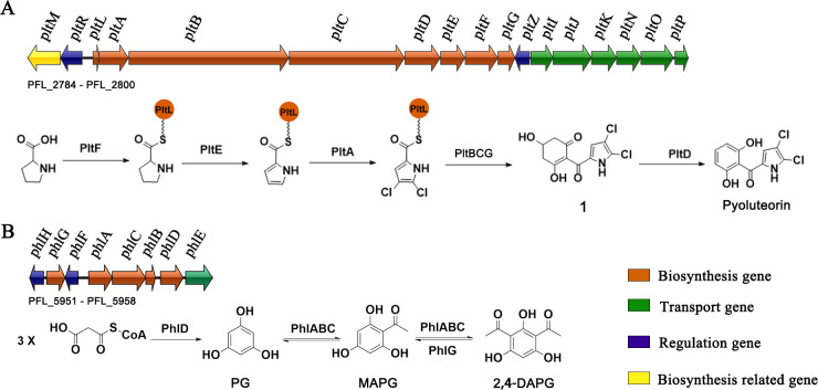
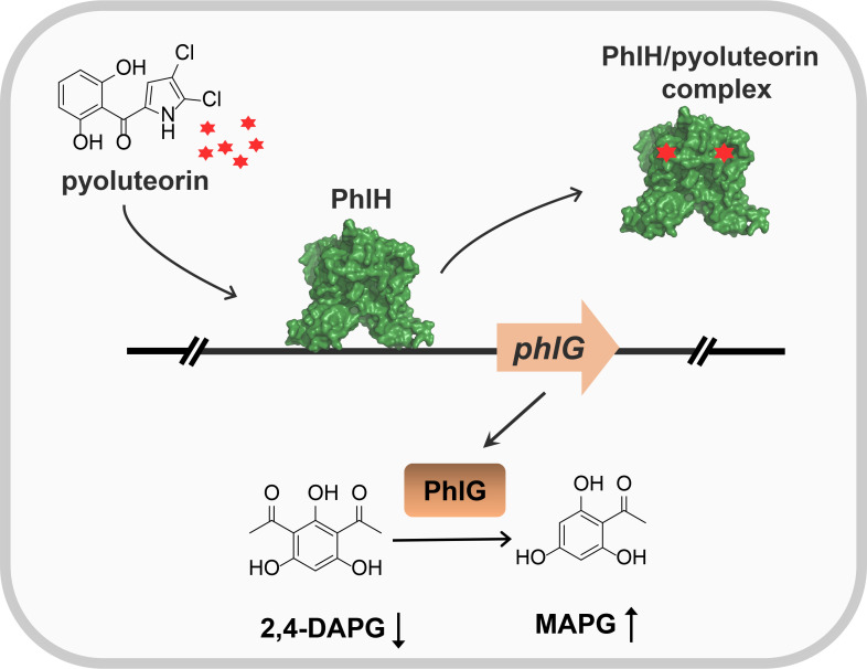
Soil and rhizosphere bacteria act as a rich source of secondary metabolites, effectively fighting against a diverse array of pathogens. Certain Pseudomonas species harbor biosynthetic gene clusters for producing both pyoluteorin and 2,4-diacetylphloroglucinol (2,4-DAPG), which are polyketides that exhibit highly similar antimicrobial spectrum against bacteria and fungi or oomycete. A complex cross talk exists between pyoluteorin and 2,4-DAPG biosynthesis, and production of 2,4-DAPG was strongly repressed by pyoluteorin, yet the underlying mechanism is still elusive. In this study, we find that the TetR family transcription factor PhlH is involved in the cross talk between pyoluteorin and 2,4-DAPG biosynthesis. PhlH binds to a palindromic sequence within the promoter of phlG (PphlG), which encodes a C-C bond hydrolase responsible for degrading 2,4-DAPG. As a signaling molecule, pyoluteorin disrupts the PhlH-PphlG complex by binding to PhlH, leading to decreased levels of 2,4-DAPG. Proteomics data suggest that pyoluteorin regulates multiple physiological processes including fatty acid biosynthesis and transportation of taurine, siderophore, and amino acids. Our work not only reveals a novel mechanism of cross talk between pyoluteorin and 2,4-DAPG biosynthesis, but also highlights pyoluteorin's role as a messenger in the complex communication network of Pseudomonas.IMPORTANCEAntibiosis serves as a crucial defense mechanism for microbes against invasive bacteria and resource competition. These bacteria typically orchestrate the production of multiple antibiotics in a coordinated fashion, wherein the synthesis of one antibiotic inhibits the generation of another. This strategic coordination allows the bacterium to focus its resources on producing the most advantageous antibiotic under specific circumstances. However, the underlying mechanisms of distinct antibiotic production in bacterial cells remain largely elusive. In this study, we reveal that the TetR family transcription factor PhlH detects the secondary metabolite pyoluteorin and mediates the cross talk between pyoluteorin and 2,4-DAPG biosynthesis in the biocontrol strain Pseudomonas protegens Pf-5. These findings hold promise for future research, potentially informing the manipulation of these systems to enhance the effectiveness of biocontrol agents.

Keywords: 2,4-DAPG; PhlH; Pseudomonas protegens; cross talk; pyoluteorin.


Conflict of interest statement

The authors declare no conflict of interest.

Figures

Fig 1
Fig 1
The biosynthetic pathways of pyoluteorin (A) and 2,4-DAPG (B) in P. protegens Pf-5. The genomic context of the BGCs for pyoluteorin spans from PFL_2784 to PFL_2800, and for 2,4-DAPG extending from PFL_5951 to PFL_5958 in P. protegens Pf-5. 3X, three molecules of malonyl-CoA. PG, phloroglucinol. MAPG, monoacetylphloroglucinol.
Fig 2
Fig 2
Comparative proteomic analysis of P. protegens Pf-5 grown with or without 20-µM pyoluteorin. (A) A volcano plot showing protein expression ratios of 20-µM pyoluteorin treated vs untreated bacterial cells. The red and blue spots indicate the upregulated and downregulated proteins, respectively. The enlarged dots represent the proteins that were mentioned in the text. (B) Top 10 hallmark functional categories highlighting the differentially expressed protein categories between P. protegens Pf-5 treated with or without 20-µM pyoluteorin. The x-axis is the enrichment factor, the spot size represents the protein number, and the spot color denotes the P value. (C) Top 10 hallmark pathways highlighting the differentially expressed protein pathways between P. protegens Pf-5 treated with or without 20-µM pyoluteorin. The x-axis is the count, and the color denotes the P value.
Fig 3
Fig 3
PhlH and PltZ interact with PphlG. (A) Multiple sequence alignment of Pf-5PhlH, Pf-5PltZ, and 2P24PhlH. The secondary structure information of Pf-5PhlH was retrieved from the crystal structure of Pf-5PhlH in complex with 2,4-DAPG (PDB code 7E1N). The strictly conserved residue for ligand binding was marked as black triangles, and unconserved residues were marked as green triangles. (B) The phlG promoter region of P. protegens Pf-5 was predicted by the BPROM program. Black lines indicate the -35 and -10 promoter regions of phlG. The binding motif of PhlH and PltZ is shown in red. The DNA probe containing the protein binding motif (PphlG F0) used for EMSA is boxed. Black arrows denote the translation start sites of phlG and phlH, respectively. EMSA of PphlG with PhlH (C) and PltZ (D). EMSA of PphlG F0 with PhlH (E) and PltZ (F). The concentration of DNA was 20 ng. The concentrations of proteins are labeled at the top of each lane.
Fig 4
Fig 4
PhlH and PltZ regulate the expression of phlG. (A) The β-galactosidase activities of the pRG970-PphlG plasmid were evaluated in wild-type (wt), ΔpltZ, ΔphlH, and ΔpltZΔphlH strains to assess the expression of phlG. (B) The transcription level of phlG was measured in the following strains: wt [wt (vector)]; mutant strains including ΔpltZ (vector), ΔphlH (vector), and ΔpltZΔphlH (vector); and complemented strains including ΔpltZ (pltZ), ΔphlH (phlH), ΔpltZΔphlH (phlH), and ΔpltZΔphlH (pltZ) via qRT-PCR assays. The expression level of zwf gene was used for normalization, and 2-ΔΔCt method was used for data analysis. (C) 2,4-DAPG production in wt [wt (vector)]; mutant strains [ΔpltZ (vector), ΔphlH (vector), and ΔpltZΔphlH (vector)]; and complemented strains [ΔpltZ (pltZ), ΔphlH (phlH), ΔpltZΔphlH (phlH), and ΔpltZΔphlH (pltZ)]. The vector in parentheses of above strains means that the strain contains empty vector pBBR1MCS-5; the pltZ or phlH in parentheses means that the strain is complemented by plasmid-borne phlH or pltZ, respectively. Error bars denote standard deviations (n = 3), P < 0.001 is displayed as ***, P < 0.01 is displayed as **, and P < 0.05 is displayed as *.
Fig 5
Fig 5
Pyoluteorin disrupts the PphlG/PhlH and PphlG/PltZ complexes. Interaction between pyoluteorin and PhlH (A) and PltZ (B) was evaluated by ITC. Data were analyzed using the MicroCal PEAQ-ITC software. Different concentrations of pyoluteorin were added to explore the stability of the PphlG/PhlH complex (C) and PphlG/PltZ complex using EMSA. The concentrations of DNA and protein were 20 ng and 0.5 µM, respectively. The concentrations of ligand are labeled at the top of each lane.
Fig 6
Fig 6
Pyoluteorin decreases 2,4-DAPG production. (A) The β-galactosidase activities of the pRG970-PphlG plasmid were evaluated in wt, ΔpltZ, ΔphlH, and ΔpltZΔphlH strains to assess the expression of phlG in the presence of 20-µM pyoluteorin or not. (B) qRT-PCR assays on phlG gene of the wt, ΔpltZ, ΔphlH, and ΔpltZΔphlH strains grown with 20-µM pyoluteorin treated or untreated culture. The expression level of zwf gene was used for normalization, and 2-ΔΔCt method was used for data analysis. (C) 2,4-DAPG production of wt, ΔpltZ, ΔphlH, and ΔpltZΔphlH strains grown with or without 20-µM pyoluteorin. (D) 2,4-DAPG production of wt strain and ΔphlG strain grown in NBGly medium. Error bars denote standard deviations (n = 3), P < 0.001 is displayed as ***, P < 0.01 is displayed as **, and P < 0.05 is displayed as *.
Fig 7
Fig 7
Comparative proteomic analysis of P. protegens Pf-5 wild-type and ΔphlG strains. (A) A volcano plot showing the differentially expressed protein in the ΔphlG strain compared with the wild-type strain of P. protegens Pf-5. For each protein, the -log10 (P value) is plotted against its log2 (fold change). The red and blue spots indicate the upregulated and downregulated proteins, respectively. The enlarged dots represent the proteins that were mentioned in the text. (B) Top 10 hallmark functional categories highlighting the differentially expressed protein categories between the ΔphlG strain and the wild-type strain. The x-axis is the enrichment factor, the spot size represents the protein number, and the spot color denotes the P value. (C) Top 10 hallmark pathways highlighting the differentially expressed protein pathways between the ΔphlG strain and the wild-type strain. The x-axis is the count, and the color denotes the P value. (D) The STRING interaction network analysis of differentially expressed proteins in the ΔphlG strain compared with the wild-type strain. Each node represents one protein, and the lines indicate the interaction between two proteins.
Fig 8
Fig 8
Proposed regulation mechanism. Pyoluteorin was sensed by the TetR-type transcriptional factor PhlH. Upon pyoluteorin signal perception, PhlH de-represses the transcription of the 2,4-DAPG hydrolase PhlG, which leads to 2,4-DAPG hydrolysis. Pyoluteorin can act as a signaling molecule to de-repress the expression of phlG.

Similar articles

Cited by

References

    1. Chater KF. 2006. Streptomyces inside-out: a new perspective on the bacteria that provide us with antibiotics. Philos Trans R Soc Lond B Biol Sci 361:761–768. doi:10.1098/rstb.2005.1758 - DOI - PMC - PubMed
    1. Alduina R, Tocchetti A, Costa S, Ferraro C, Cancemi P, Sosio M, Donadio S. 2020. A Two-component regulatory system with opposite effects on glycopeptide antibiotic biosynthesis and resistance. Sci Rep 10:6200. doi:10.1038/s41598-020-63257-4 - DOI - PMC - PubMed
    1. Donia MS, Cimermancic P, Schulze CJ, Wieland Brown LC, Martin J, Mitreva M, Clardy J, Linington RG, Fischbach MA. 2014. A systematic analysis of biosynthetic gene clusters in the human microbiome reveals a common family of antibiotics. Cell 158:1402–1414. doi:10.1016/j.cell.2014.08.032 - DOI - PMC - PubMed
    1. Skinnider MA, Johnston CW, Gunabalasingam M, Merwin NJ, Kieliszek AM, MacLellan RJ, Li H, Ranieri MRM, Webster ALH, Cao MPT, Pfeifle A, Spencer N, To QH, Wallace DP, Dejong CA, Magarvey NA. 2020. Comprehensive prediction of secondary metabolite structure and biological activity from microbial genome sequences. Nat Commun 11:6058. doi:10.1038/s41467-020-19986-1 - DOI - PMC - PubMed
    1. Haas D, Défago G. 2005. Biological control of soil-borne pathogens by fluorescent pseudomonads. Nat Rev Microbiol 3:307–319. doi:10.1038/nrmicro1129 - DOI - PubMed

Publication types

MeSH terms

Supplementary concepts