Review
. 2024 Mar;310(3):e231220.
doi: 10.1148/radiol.231220.

Interpretation, Reporting, and Clinical Applications of Liver MR Elastography

Affiliations
Review

Interpretation, Reporting, and Clinical Applications of Liver MR Elastography

Guilherme Moura Cunha et al. Radiology. 2024 Mar.

Abstract

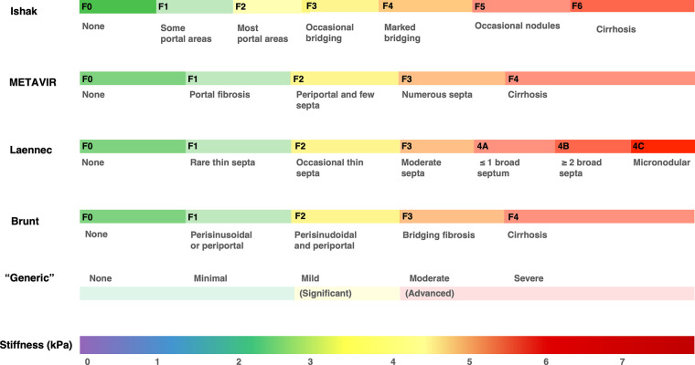
Chronic liver disease is highly prevalent and often leads to fibrosis or cirrhosis and complications such as liver failure and hepatocellular carcinoma. The diagnosis and staging of liver fibrosis is crucial to determine management and mitigate complications. Liver biopsy for histologic assessment has limitations such as sampling bias and high interreader variability that reduce precision, which is particularly challenging in longitudinal monitoring. MR elastography (MRE) is considered the most accurate noninvasive technique for diagnosing and staging liver fibrosis. In MRE, low-frequency vibrations are applied to the abdomen, and the propagation of shear waves through the liver is analyzed to measure liver stiffness, a biomarker for the detection and staging of liver fibrosis. As MRE has become more widely used in clinical care and research, different contexts of use have emerged. This review focuses on the latest developments in the use of MRE for the assessment of liver fibrosis; provides guidance for image acquisition and interpretation; summarizes diagnostic performance, along with thresholds for diagnosis and staging of liver fibrosis; discusses current and emerging clinical applications; and describes the latest technical developments.


Conflict of interest statement

Disclosures of conflicts of interest: G.M.C. No relevant relationships. B.F. No relevant relationships. P.J.N. No relevant relationships. D.O. No relevant relationships. S.K.V. No relevant relationships. R.L.E. Research contract from Resoundant, royalties paid to institution (Mayo Clinic) from Resoundant, patents owned by institution (Mayo Clinic), and stock or stock options from Resoundant; the Mayo Clinic and R.L.E. have intellectual property rights and a financial interest in MR elastographic technology. C.B.S. Research grants from American College of Radiology, Bayer, Foundation for the National Institutes of Health, General Electric, Gilead, Pfizer, Philips, Siemens, and V Foundation; lab service agreements with OrsoBio, Enanta Pharmaceuticals, Gilead, ICON, Intercept Pharmaceuticals, NuSirt BioPharma, Shire, Synageva, and Takeda; institutional consulting for Bristol Myers Squibb, Exact Sciences, IBM Watson, and Pfizer; personal consulting for Altimmune, Ascelia Pharma, Blade Therapeutics, Boehringer Ingelheim, Epigenomics, and Guerbet; receipt of royalties and/or honoraria from Medscape and Wolters Kluwer; ownership of stock options in Livivos; unpaid advisory board position for Quantix Bio; and chief medical officer for Livivos (unsalaried position with stock options and stock) through June 28, 2023, and currently principal advisor for Livivos (both appointments approved by his university). A.T. Steering committee member for Liver Imaging Reporting and Data System.

Figures

None
Graphical abstract
RSNA Quantitative Imaging Biomarkers Alliance recommendations for
region of interest placement and measurement of shear-wave propagation in MR
elastography. A region of interest (outline) is drawn on the (A) axial
source magnitude image, with simultaneous visualization of the (B) wave
image and the (C) elastogram with overlaid confidence map (hatching), in
such a way as to include the largest portion of liver tissue. The region of
interest should be placed on individual image sections excluding areas of
low wave propagation, large vessels, focal lesions, marginal liver tissues
(to avoid edge effect), and areas identified by the inversion algorithm as
invalid (hatching in C). The mean liver stiffness is reported by recording
the mean stiffness value of each region of interest (ie, each section) and
then calculating the mean value for all sections, weighted by the size of
the region of interest. In the wave image (B), red indicates peaks and blue
indicates troughs. Highly saturated colors indicate high wave amplitude, and
black indicates low wave amplitude. Color bar in the elastogram (C)
indicates stiffness range.
Figure 1:
RSNA Quantitative Imaging Biomarkers Alliance recommendations for region of interest placement and measurement of shear-wave propagation in MR elastography. A region of interest (outline) is drawn on the (A) axial source magnitude image, with simultaneous visualization of the (B) wave image and the (C) elastogram with overlaid confidence map (hatching), in such a way as to include the largest portion of liver tissue. The region of interest should be placed on individual image sections excluding areas of low wave propagation, large vessels, focal lesions, marginal liver tissues (to avoid edge effect), and areas identified by the inversion algorithm as invalid (hatching in C). The mean liver stiffness is reported by recording the mean stiffness value of each region of interest (ie, each section) and then calculating the mean value for all sections, weighted by the size of the region of interest. In the wave image (B), red indicates peaks and blue indicates troughs. Highly saturated colors indicate high wave amplitude, and black indicates low wave amplitude. Color bar in the elastogram (C) indicates stiffness range.
Example axial T2-weighted (T2 W) MRI scans (left), wave images
(middle), and elastograms (right) illustrating the contexts of use of MR
elastography in patients with advanced fibrosis with different liver disease
etiologies. The reported stiffness threshold for fibrosis stages may vary
based on differences in study population and reference standard (ie,
histologic examination) sampling variability. Based on consensus
interpretation of multiple studies and meta-analysis, and to simplify the
interpretation of MR elastographic stiffness in clinical care, a common set
of simple thresholds is recommended regardless of the underlying etiology of
chronic liver disease: 3.0 kPa for stage F1 or higher, 3.5 kPa for stage F2
or higher, 4.0 kPa for stage F3 or higher, and 5.0 kPa for stage F4. HBV =
hepatitis B virus infection, HCV = hepatitis C virus infection, MASH =
metabolic dysfunction–associated steatohepatitis, PSC = primary
sclerosing cholangitis.
Figure 2:
Example axial T2-weighted (T2 W) MRI scans (left), wave images (middle), and elastograms (right) illustrating the contexts of use of MR elastography in patients with advanced fibrosis with different liver disease etiologies. The reported stiffness threshold for fibrosis stages may vary based on differences in study population and reference standard (ie, histologic examination) sampling variability. Based on consensus interpretation of multiple studies and meta-analysis, and to simplify the interpretation of MR elastographic stiffness in clinical care, a common set of simple thresholds is recommended regardless of the underlying etiology of chronic liver disease: 3.0 kPa for stage F1 or higher, 3.5 kPa for stage F2 or higher, 4.0 kPa for stage F3 or higher, and 5.0 kPa for stage F4. HBV = hepatitis B virus infection, HCV = hepatitis C virus infection, MASH = metabolic dysfunction–associated steatohepatitis, PSC = primary sclerosing cholangitis.
Graph shows reported MR elastographic (MRE) stiffness thresholds (in
kilopascals) for fibrosis staging. Dotted lines represent the thresholds
recommended by the Liver Imaging Reporting and Data System Quantitative
Imaging Working Group: 3.0 kPa or higher for stage F1 (mild fibrosis), 3.5
kPa or higher for stage F2 (significant fibrosis), 4.0 kPa or higher for
stage F3 (advanced fibrosis), and 5.0 kPa or higher for stage F4
(cirrhosis). * = These studies report results for different
sequences, frequencies, readers, or stiffness thresholds investigated.
† = “Various liver diseases” includes hepatitis B virus
infection, hepatitis C virus infection, metabolic
dysfunction–associated steatohepatitis (MASH), and autoimmune
hepatitis. MAFLD = metabolic dysfunction–associated fatty liver
disease.
Figure 3:
Graph shows reported MR elastographic (MRE) stiffness thresholds (in kilopascals) for fibrosis staging. Dotted lines represent the thresholds recommended by the Liver Imaging Reporting and Data System Quantitative Imaging Working Group: 3.0 kPa or higher for stage F1 (mild fibrosis), 3.5 kPa or higher for stage F2 (significant fibrosis), 4.0 kPa or higher for stage F3 (advanced fibrosis), and 5.0 kPa or higher for stage F4 (cirrhosis). * = These studies report results for different sequences, frequencies, readers, or stiffness thresholds investigated. † = “Various liver diseases” includes hepatitis B virus infection, hepatitis C virus infection, metabolic dysfunction–associated steatohepatitis (MASH), and autoimmune hepatitis. MAFLD = metabolic dysfunction–associated fatty liver disease.
Graph shows multiple scoring systems that have been proposed for
staging liver fibrosis. These systems differ in their purpose, definitions
of fibrosis stages, and subgroups. The use of liver stiffness as a
continuous variable has advantages over ordinal categories. It may provide
better clinical and prognostic correlation, especially given that most
staging systems group patients with cirrhosis in a single category, without
taking into consideration the severity of cirrhosis or any longitudinal
histologic progression or regression. Brunt = Brunt scoring system, F =
fibrosis stage, Ishak = Ishak score, Laennec = Laennec staging system,
METAVIR = Meta-analysis of Histological Data in Viral Hepatitis scoring
system.
Figure 4:
Graph shows multiple scoring systems that have been proposed for staging liver fibrosis. These systems differ in their purpose, definitions of fibrosis stages, and subgroups. The use of liver stiffness as a continuous variable has advantages over ordinal categories. It may provide better clinical and prognostic correlation, especially given that most staging systems group patients with cirrhosis in a single category, without taking into consideration the severity of cirrhosis or any longitudinal histologic progression or regression. Brunt = Brunt scoring system, F = fibrosis stage, Ishak = Ishak score, Laennec = Laennec staging system, METAVIR = Meta-analysis of Histological Data in Viral Hepatitis scoring system.
Example of the MR elastography (MRE) plus Fibrosis-4 (FIB-4) (MEFIB)
sequential approach used to rule in or rule out significant fibrosis in a
66-year-old man with clinical suspicion of significant fibrosis. A FIB-4
value of 5.15 (higher than the ≥1.6 threshold) computed from age,
aspartate aminotransferase, alanine transaminase, and platelet count
justified proceeding to MRE, which revealed a mean stiffness of 5.7 kPa. The
axial elastogram represents a stiffness map expressed in kilopascals, on a
scale from 0 to 8 kPa, where low stiffness is represented in purple and high
stiffness in red, with a confidence mask overlaid (hatching). The
combination of a FIB-4 value of 1.6 or greater and an MRE-derived liver
stiffness of 3.3 kPa or greater has a high positive predictive value to rule
in significant fibrosis (≥F2). A liver biopsy performed in a research
setting confirmed cirrhosis (F4) related to metabolic
dysfunction–associated steatohepatitis.
Figure 5:
Example of the MR elastography (MRE) plus Fibrosis-4 (FIB-4) (MEFIB) sequential approach used to rule in or rule out significant fibrosis in a 66-year-old man with clinical suspicion of significant fibrosis. A FIB-4 value of 5.15 (higher than the ≥1.6 threshold) computed from age, aspartate aminotransferase, alanine transaminase, and platelet count justified proceeding to MRE, which revealed a mean stiffness of 5.7 kPa. The axial elastogram represents a stiffness map expressed in kilopascals, on a scale from 0 to 8 kPa, where low stiffness is represented in purple and high stiffness in red, with a confidence mask overlaid (hatching). The combination of a FIB-4 value of 1.6 or greater and an MRE-derived liver stiffness of 3.3 kPa or greater has a high positive predictive value to rule in significant fibrosis (≥F2). A liver biopsy performed in a research setting confirmed cirrhosis (F4) related to metabolic dysfunction–associated steatohepatitis.
Example of the MRI–aspartate aminotransferase (AST) (MAST)
combined approach used to rule in or rule out metabolic
dysfunction–associated steatohepatitis (MASH) in a 52-year-old woman
with clinical suspicion of MASH. The MAST score is computed from MR
elastography (MRE)–derived stiffness, MRI proton density fat fraction
(PDFF), and AST. (A) The axial elastogram represents a stiffness map
expressed in kilopascals, on a scale from 0 to 8 kPa, where low stiffness is
represented in purple and high stiffness in red, with a confidence mask
overlaid (hatching). The mean MRE stiffness was 2.7 kPa. (B) MRI proton
density fat fraction, determined from the axial MRI scan, was 20.5%,
indicating moderate to severe steatosis. With an AST value of 144 U/L, the
computed MAST score was 0.332. A value of 0.242 or higher is used to rule in
fibro-MASH (MASH activity score of 4 or greater and fibrosis stage of F2 or
greater). A liver biopsy performed in a research setting confirmed a MASH
activity score of 5 and a fibrosis stage of F2.
Figure 6:
Example of the MRI–aspartate aminotransferase (AST) (MAST) combined approach used to rule in or rule out metabolic dysfunction–associated steatohepatitis (MASH) in a 52-year-old woman with clinical suspicion of MASH. The MAST score is computed from MR elastography (MRE)–derived stiffness, MRI proton density fat fraction (PDFF), and AST. (A) The axial elastogram represents a stiffness map expressed in kilopascals, on a scale from 0 to 8 kPa, where low stiffness is represented in purple and high stiffness in red, with a confidence mask overlaid (hatching). The mean MRE stiffness was 2.7 kPa. (B) MRI proton density fat fraction, determined from the axial MRI scan, was 20.5%, indicating moderate to severe steatosis. With an AST value of 144 U/L, the computed MAST score was 0.332. A value of 0.242 or higher is used to rule in fibro-MASH (MASH activity score of 4 or greater and fibrosis stage of F2 or greater). A liver biopsy performed in a research setting confirmed a MASH activity score of 5 and a fibrosis stage of F2.

References

    1. Chi H , Hansen BE , Tang WY , et al . Multiple biopsy passes and the risk of complications of percutaneous liver biopsy . Eur J Gastroenterol Hepatol 2017. ; 29 ( 1 ): 36 – 41 . - PubMed
    1. Bedossa P , Carrat F . Liver biopsy: the best, not the gold standard . J Hepatol 2009. ; 50 ( 1 ): 1 – 3 . - PubMed
    1. Bedossa P , Dargère D , Paradis V . Sampling variability of liver fibrosis in chronic hepatitis C . Hepatology 2003. ; 38 ( 6 ): 1449 – 1457 . - PubMed
    1. Goodman ZD . Grading and staging systems for inflammation and fibrosis in chronic liver diseases . J Hepatol 2007. ; 47 ( 4 ): 598 – 607 . - PubMed
    1. Yeh WC , Li PC , Jeng YM , et al . Elastic modulus measurements of human liver and correlation with pathology . Ultrasound Med Biol 2002. ; 28 ( 4 ): 467 – 474 . - PubMed

MeSH terms