. 2024 Apr 10;15(4):e0345623.
doi: 10.1128/mbio.03456-23. Epub 2024 Mar 12.

Control of three-carbon amino acid homeostasis by promiscuous importers and exporters in Bacillus subtilis: role of the "sleeping beauty" amino acid exporters

Affiliations

Control of three-carbon amino acid homeostasis by promiscuous importers and exporters in Bacillus subtilis: role of the "sleeping beauty" amino acid exporters

Robert Warneke et al. mBio. .

Abstract

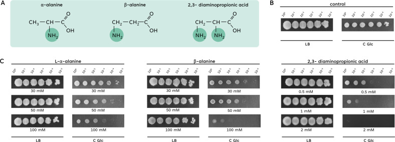
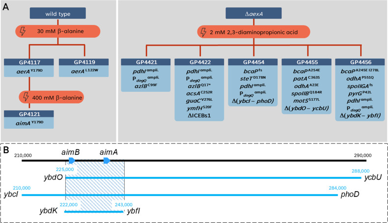
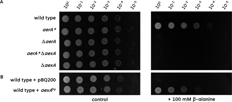
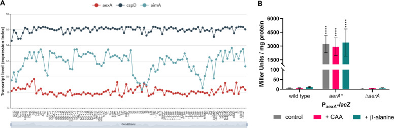
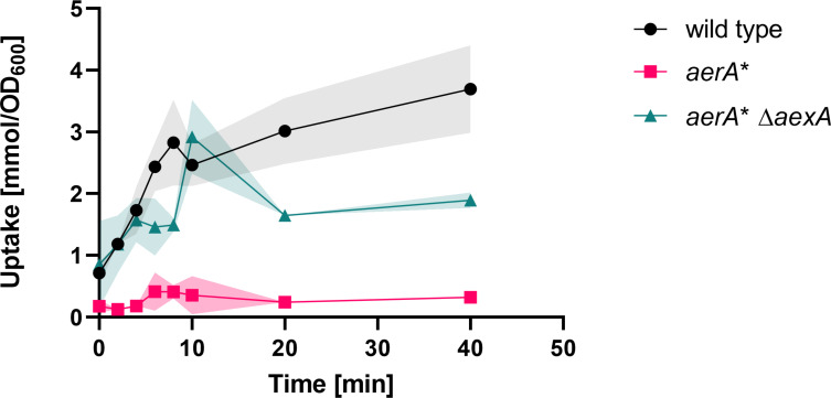
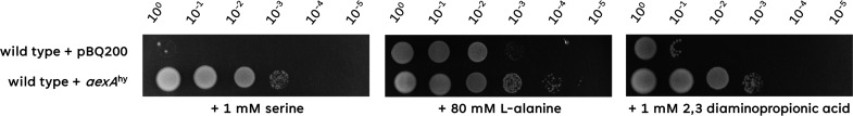
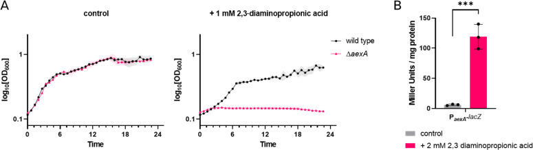
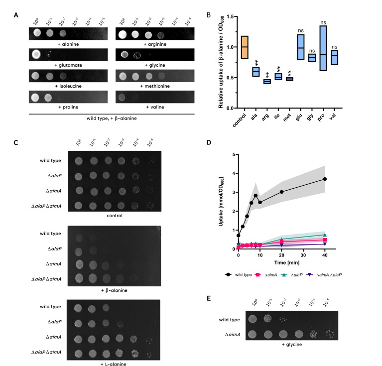
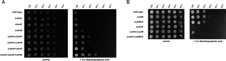
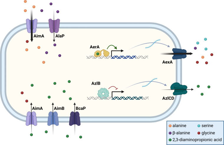
The Gram-positive model bacterium Bacillus subtilis can acquire amino acids by import, de novo biosynthesis, or degradation of proteins and peptides. The accumulation of several amino acids inhibits the growth of B. subtilis, probably due to misincorporation into cellular macromolecules such as proteins or peptidoglycan or due to interference with other amino acid biosynthetic pathways. Here, we studied the adaptation of B. subtilis to toxic concentrations of the three-carbon amino acids L-alanine, β-alanine, and 2,3-diaminopropionic acid, as well as the two-carbon amino acid glycine. Resistance to the non-proteinogenic amino acid β-alanine, which is a precursor for coenzyme A biosynthesis, is achieved by mutations that either activate a cryptic amino acid exporter, AexA (previously YdeD), or inactivate the amino acid importers AimA, AimB (previously YbxG), and BcaP. The aexA gene is very poorly expressed under most conditions studied. However, mutations affecting the transcription factor AerA (previously YdeC) can result in strong constitutive aexA expression. AexA is the first characterized member of a group of amino acid exporters in B. subtilis, which are all very poorly expressed. Therefore, we suggest to call this group "sleeping beauty amino acid exporters." 2,3-Diaminopropionic acid can also be exported by AexA, and this amino acid also seems to be a natural substrate of AerA/AexA, as it can cause a slight but significant induction of aexA expression, and AexA also provides some natural resistance toward 2,3-diaminopropionic acid. Moreover, our work shows how low-specificity amino acid transporters contribute to amino acid homeostasis in B. subtilis.IMPORTANCEEven though Bacillus subtilis is one of the most-studied bacteria, amino acid homeostasis in this organism is not fully understood. We have identified import and export systems for the C2 and C3 amino acids. Our work demonstrates that the responsible amino acid permeases contribute in a rather promiscuitive way to amino acid uptake. In addition, we have discovered AexA, the first member of a group of very poorly expressed amino acid exporters in B. subtilis that we call "sleeping beauty amino acid exporters." The expression of these transporters is typically triggered by mutations in corresponding regulator genes that are acquired upon exposure to toxic amino acids. These exporters are ubiquitous in all domains of life. It is tempting to speculate that many of them are not expressed until the cells experience selective pressure by toxic compounds, and they protect the cells from rare but potentially dangerous encounters with such compounds.

Keywords: Bacillus subtilis; amino acid export; amino acid uptake; diaminopropionic acid; sleeping beauty amino acid exporters; β-alanine.


Conflict of interest statement

The authors declare no conflict of interest.

Figures

Fig 1
Fig 1
Growth of B. subtilis in the presence of alanine derivates. (A) The three alanine derivates, α-alanine, β-alanine, and 2,3-diaminopropionic acid, are displayed. The position of the amino group is highlighted. (B and C) The growth of the wild-type strain 168 was compared on complex medium (LB) and minimal medium (C Glc) in the absence or presence of the alanine derivates L-α-alanine, β-alanine, and 2,3-diaminopropionic acid. The cells were grown in C-Glc minimal medium or LB complex medium to an OD600 of 1.0, and serial dilutions (10-fold) were prepared. These samples were plated on C-Glc minimal plates or LB plates containing 30 mM, 50 mM, or 100 mM L-α-alanine or β-alanine, or 0.5–2 mM 2,3-diaminopropionic acid. The plates were incubated at 37°C for 48 h.
Fig 2
Fig 2
A suppressor screen in the presence of alanine derivates. (A) Evolutionary trajectory of the wild-type strain exposed to toxic concentrations of β-alanine or the ΔaexA mutant exposed to toxic concentrations of 2,3-diaminopropionic acid. (B) Genetic regions affected by deletions in the suppressor strains (GP4454, GP4455, and GP4456) under 2,3-diaminopropionic acid stress. Each strain’s deleted regions are demarcated, and an overlay highlights the shared deleted region, denoted by a blue-striped rectangle. This shared deleted region suggests a common genetic adaptation in response to the specific stress condition.
Fig 3
Fig 3
Overexpression of aexA is required for β-alanine resistance. (A) The sensitivity of the wild type, aerA*, ΔaerA, aerA*ΔaexA, and ΔaexA mutant to β-alanine was tested. The cells were grown in C-Glc minimal medium to an OD600 of 1.0, and serial dilutions (10-fold) were prepared. These samples were plated on C-Glc minimal plates containing no β-alanine or 100 mM β-alanine and incubated at 37°C for 48 h. (B) The sensitivity of the wild type harboring either the empty vector pBQ200 or pGP3727 (aexAhy) to β-alanine was tested. The cells were grown in C-Glc minimal medium (with erythromycin and lincomycin) to an OD600 of 1.0, and serial dilutions (10-fold) were prepared. These samples were plated on C-Glc minimal plates containing no β-alanine or 100 mM β-alanine and incubated at 37°C for 48 h.
Fig 4
Fig 4
The aexA gene is only expressed in the aerA* mutant. A. The Expression Browser of SubtiWiki allows a direct comparison of the expression levels of two or more genes (here aexA, cspD, and aimA). The expression level of cspD serves as an example of a gene with high constitutive expression. For aimA, a moderate expression is observed, while the aexA gene is virtually never expressed. For details on the conditions displayed on the X-axis, please consult the interactive expression browser of the SubtiWiki database (http://www.subtiwiki.uni-goettingen.de/v4/expression?gene=3BD05DE3ED5B293980F47EB621C54C7339F78DC7, 29). (B) The expression of the aexA promoter was monitored in strains that harbor the aexA-lacZ reporter gene fusion integrated into the chromosomal amyE gene. The tested strains were wild type, the aerA* mutant, as well as the deletion mutant ΔaerA. Cultures were grown in C-Glc minimal medium supplemented with or without casaminoacids (CAA; magenta) or β-alanine (cyan) to the early exponential phase (OD578 of about 0.6–0.8) and then harvested for β-galactosidase enzyme activity assays. The values for the β-galactosidase activity indicated for each strain represent three independently grown cultures, and for each sample, enzyme activity was determined twice.
Fig 5
Fig 5
Intracellular accumulation of radiolabeled β-alanine. For the quantification of intracellular accumulation of [1–14C]β-alanine, cells of the wild type (black circles), aerA* (magenta squares), and aerA* ΔaexA (cyan triangles) mutant were cultivated in C-Glc minimal medium to an OD600 of approximately 0.8. The cells were then diluted with C-Glc minimal medium containing [1–14C]β-alanine to reach a final concentration of 30 mM. The accumulation of intracellular [1–14C]β-alanine was assayed after 0, 2, 4, 6, 8, 10, 20, and 40 minutes. n = 3.
Fig 6
Fig 6
Overexpression of AexA confers resistance against L-serine, L-alanine, and 2,3-diaminopropionic acid. The sensitivity of the wild type harboring the empty vector pBQ200 or pGP3727 aexAhy to L-serine, L-alanine, or 2,3-diaminopropionic acid was tested. The cells were grown in C-Glc minimal medium to an OD600 of 1.0, and serial dilutions (10-fold) were prepared. These samples were plated on C-Glc minimal plates containing 1 mM L-serine, 80 mM L-alanine, or 1 mM 2,3-diaminopropionic acid and incubated at 37°C for 48 h.
Fig 7
Fig 7
2,3-Diaminopropionic acid is a weak inducer of aexA expression. (A) The growth of the wild type and the ΔaexA mutant (GP3955) on C Glc minimal medium was tested in the presence or absence of 1 mM 2,3-diaminopropionic acid. (B) The expression of the aexA promoter was monitored in strains that harbor the aexA-lacZ reporter gene fusion integrated into the chromosomal amyE gene. Cultures were grown in C Glc minimal medium in the presence or absence of 2 mM 2,3-diaminopropionic acid to the early exponential phase (OD578 of about 0.6–0.8) and then harvested for β-galactosidase enzyme activity assays. The values for the β-galactosidase activity indicated for each strain represent three independently grown cultures, and for each sample, enzyme activity was determined twice.
Fig 8
Fig 8
The presence of different amino acids can prevent β-alanine toxicity. (A) The growth of the wild-type strain in the presence of β-alanine and either alanine, arginine, isoleucine, methionine, glutamate, glycine, proline, or valine was checked. The cells were grown in C-Glc minimal medium to an OD600 of 1.0, and serial dilutions (10-fold) were prepared. These samples were plated on C-Glc minimal plates containing 100 mM β-alanine together with 30 mM of the respective amino acid and incubated at 37°C for 48 h. (B) To check the relative uptake of β-alanine in the presence or absence of the tested amino acid, the wild type was grown in C-Glc minimal medium containing 30 mM β-alanine spiked with radiolabeled [1–14C]β-alanine with or without 5 mM of the respective amino acid to an OD600 of approximately 0.8. The accumulation of intracellular [1–14C]β-alanine was assayed after 40 minutes. n = 3 (C) The sensitivity of the wild type 168, ΔalaP (GP4136), ΔaimA (GP3590), and ΔalaP ΔaimA (GP4135) to β-alanine or L-alanine was tested. The cells were grown in C-Glc minimal medium to an OD600 of 1.0, and serial dilutions (10-fold) were prepared. These samples were plated on C-Glc minimal plates containing 100 mM β-alanine or L-alanine and incubated at 37°C for 48 h. (D) For the quantification of intracellular accumulation of [1–14C]β-alanine, cells of the wild type (168, black circles), ΔalaP (GP4136, cyan triangles pointing upward), ΔaimA (GP3590, magenta squares), and ΔalaP ΔaimA (GP4135, purple triangles pointing downward) mutant were cultivated in C-Glc minimal medium to an OD600 of approximately 0.8. The cells were then diluted with C-Glc minimal medium containing [1–14C]β-alanine to reach a final concentration of 30 mM. The accumulation of intracellular [1–14C]β-alanine was assayed after 0, 2, 4, 6, 8, 10, 20, and 40 minutes. n = 3 (E) The sensitivity of the wild type 168 and the ΔaimA (GP3590) to glycine was tested. The cells were grown in C-Glc minimal medium to an OD600 of 1.0, and serial dilutions (10-fold) were prepared. These samples were plated on C-Glc minimal plates containing 200 mM glycine and incubated at 37°C for 48 h.
Fig 9
Fig 9
Sensitivity of mutant strains to 2,3-diaminiopropionic acid. (A) The sensitivity of the wild type (168), ΔaimA (GP2786), ΔbcaP (GP4484), ΔaimB (GP2396), ΔaimA ΔbcaP (GP2949), ΔaimA ΔaimB (GP2951), ΔaimB ΔbcaP (GP2952), and ΔaimA ΔaimB ΔbcaP (GP2950) to 2,3-diaminopropionic acid was tested. The cells were grown in C-Glc minimal medium to an OD600 of 1.0, and serial dilutions (10-fold) were prepared. These samples were plated on C-Glc minimal plates containing no, 100 mM, or 200 mM β-alanine and incubated at 37°C for 48 h. (B) The sensitivity of the wild type (168), ΔazlB (GP3600), ΔazlBCD (GP3623), ΔaexA (GP3955), ΔaexA ΔazlB (GP4384), and ΔaexA ΔazlBCD (GP4385) to 2,3-diaminopropionic acid was tested. The cells were grown in C-Glc minimal medium to an OD600 of 1.0, and serial dilutions (10-fold) were prepared. These samples were plated on C-Glc minimal plates containing no, 100 mM, or 200 mM β-alanine and incubated at 37°C for 48 h.
Fig 10
Fig 10
Transport of C2 and C3 amino acids in B. subtilis. The uptake and export systems for the C2 and C3 amino acids are shown in the left and right parts of the figure, respectively. The amino acid exporters AexA and AzlCD are only expressed if the transcription factors AerA and AzlB have acquired mutations that allow their DNA binding in the absence of the inducer or in the presence of 2,3-diaminopropionic acid (for AerA).

Similar articles

Cited by

References

    1. Egan AJF, Errington J, Vollmer W. 2020. Regulation of peptidoglycan synthesis and remodelling. Nat Rev Microbiol 18:446–460. doi:10.1038/s41579-020-0366-3 - DOI - PubMed
    1. Brown GM, Reynolds JJ. 1963. Biogenesis of the water-soluble vitamins. Annu Rev Biochem 32:419–462. doi:10.1146/annurev.bi.32.070163.002223 - DOI - PubMed
    1. Begley TP, Kinsland C, Strauss E. 2001. The biosynthesis of coenzyme A in bacteria. Vitam Horm 61:157–171. doi:10.1016/s0083-6729(01)61005-7 - DOI - PubMed
    1. Calogero S, Gardan R, Glaser P, Schweizer J, Rapoport G, Debarbouille M. 1994. RocR, a novel regulatory protein controlling arginine utilization in Bacillus subtilis, belongs to the NtrC/NifA family of transcriptional activators. J Bacteriol 176:1234–1241. doi:10.1128/jb.176.5.1234-1241.1994 - DOI - PMC - PubMed
    1. Warneke R, Garbers TB, Herzberg C, Aschenbrandt G, Ficner R, Stülke J. 2023. Ornithine is the central intermediate in the arginine degradative pathway and its regulation in Bacillus subtilis. J Biol Chem 299:104944. doi:10.1016/j.jbc.2023.104944 - DOI - PMC - PubMed

MeSH terms

LinkOut - more resources