. 2024 Mar 12;134(8):e178263.
doi: 10.1172/JCI178263.

Neutralizing IFN-γ autoantibodies are rare and pathogenic in HLA-DRB1*15:02 or 16:02 individuals

Affiliations

Neutralizing IFN-γ autoantibodies are rare and pathogenic in HLA-DRB1*15:02 or 16:02 individuals

Jessica N Peel et al. J Clin Invest. .

Abstract

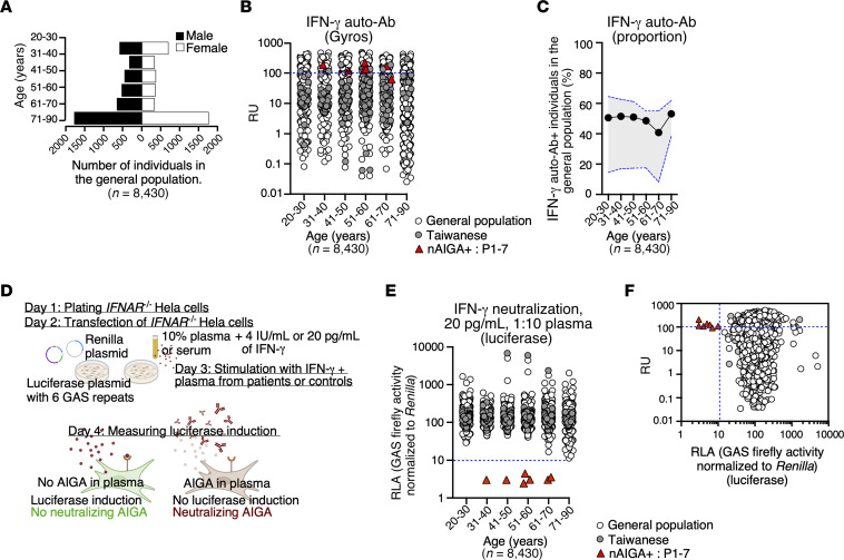
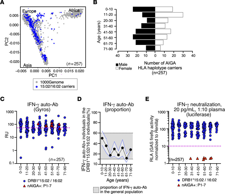
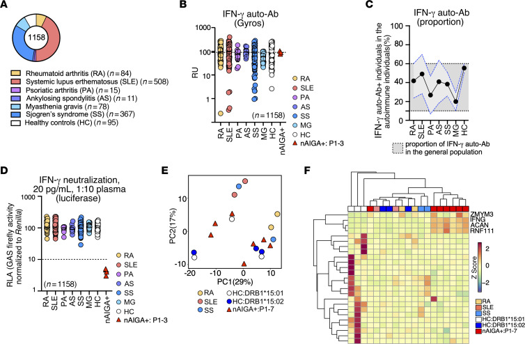
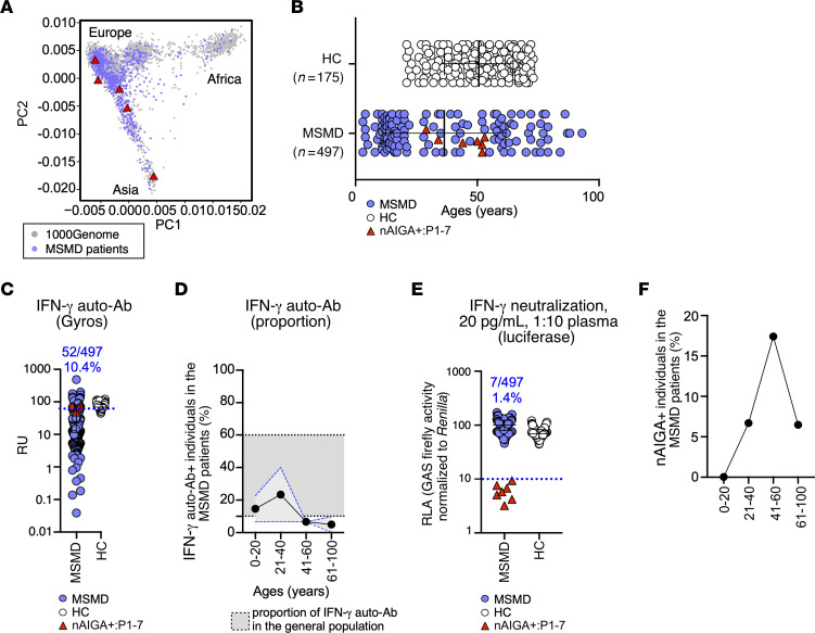
BACKGROUNDWeakly virulent environmental mycobacteria (EM) can cause severe disease in HLA-DRB1*15:02 or 16:02 adults harboring neutralizing anti-IFN-γ autoantibodies (nAIGAs). The overall prevalence of nAIGAs in the general population is unknown, as are the penetrance of nAIGAs in HLA-DRB1*15:02 or 16:02 individuals and the proportion of patients with unexplained, adult-onset EM infections carrying nAIGAs.METHODSThis study analyzed the detection and neutralization of anti-IFN-γ autoantibodies (auto-Abs) from 8,430 healthy individuals of the general population, 257 HLA-DRB1*15:02 or 16:02 carriers, 1,063 patients with autoimmune disease, and 497 patients with unexplained severe disease due to EM.RESULTSWe found that anti-IFN-γ auto-Abs detected in 4,148 of 8,430 healthy individuals (49.2%) from the general population of an unknown HLA-DRB1 genotype were not neutralizing. Moreover, we did not find nAIGAs in 257 individuals carrying HLA-DRB1* 15:02 or 16:02. Additionally, nAIGAs were absent in 1,063 patients with an autoimmune disease. Finally, 7 of 497 patients (1.4%) with unexplained severe disease due to EM harbored nAIGAs.CONCLUSIONThese findings suggest that nAIGAs are isolated and that their penetrance in HLA-DRB1*15:02 or 16:02 individuals is low, implying that they may be triggered by rare germline or somatic variants. In contrast, the risk of mycobacterial disease in patients with nAIGAs is high, confirming that these nAIGAs are the cause of EM disease.FUNDINGThe Laboratory of Human Genetics of Infectious Diseases is supported by the Howard Hughes Medical Institute, the Rockefeller University, the St. Giles Foundation, the National Institutes of Health (NIH) (R01AI095983 and U19AIN1625568), the National Center for Advancing Translational Sciences (NCATS), the NIH Clinical and Translational Science Award (CTSA) program (UL1 TR001866), the French National Research Agency (ANR) under the "Investments for the Future" program (ANR-10-IAHU-01), the Integrative Biology of Emerging Infectious Diseases Laboratory of Excellence (ANR-10-LABX-62-IBEID), ANR-GENMSMD (ANR-16-CE17-0005-01), ANR-MAFMACRO (ANR-22-CE92-0008), ANRSECTZ170784, the French Foundation for Medical Research (FRM) (EQU201903007798), the ANRS-COV05, ANR GENVIR (ANR-20-CE93-003), and ANR AI2D (ANR-22-CE15-0046) projects, the ANR-RHU program (ANR-21-RHUS-08-COVIFERON), the European Union's Horizon 2020 research and innovation program under grant agreement no. 824110 (EASI-genomics), the Square Foundation, Grandir - Fonds de solidarité pour l'enfance, the Fondation du Souffle, the SCOR Corporate Foundation for Science, the Battersea & Bowery Advisory Group, William E. Ford, General Atlantic's Chairman and Chief Executive Officer, Gabriel Caillaux, General Atlantic's Co-President, Managing Director, and Head of business in EMEA, and the General Atlantic Foundation, Institut National de la Santé et de la Recherche Médicale (INSERM) and of Paris Cité University. JR was supported by the INSERM PhD program for doctors of pharmacy (poste d'accueil INSERM). JR and TLV were supported by the Bettencourt-Schueller Foundation and the MD-PhD program of the Imagine Institute. MO was supported by the David Rockefeller Graduate Program, the Funai Foundation for Information Technology (FFIT), the Honjo International Scholarship Foundation (HISF), and the New York Hideyo Noguchi Memorial Society (HNMS).

Keywords: Autoimmune diseases; Cytokines; Genetics; Immunology.


Figures

Figure 1
Figure 1. Nonneutralizing IFN-γ auto-Abs are detected in the general population.
(A) Age and sex distribution of individuals from different cohorts are depicted in a bar plot. These cohorts include the French Blood Bank (EFS), 3C cohort, French CONSTANCES cohort, Cerba Healthcare cohort, and a cohort of Taiwanese healthy donors. (B) The detection of IFN-γ auto-Abs in the general population (white circles) compared with Taiwanese healthy donors (grey circles) and patients infected with EM and harboring nAIGAs, represented by red triangles, is shown in a dot plot. The detection thresholds are determined by auto-Abs against IFN-γ from patients with EM due to nAIGA (blue dotted line), measured in response units (RU). (C) Proportions of individuals positive for IFN-γ auto-Abs detection by Gyros are presented by decade, with standard deviation outlined in grey and a blue dotted line. (D) A schematic representation of the neutralization assay developed in IFNAR–/– HeLa cells using a luciferase system is provided. The assay involves stimulation of transfected cells with IFN-γ and measurement of luciferase induction. (E) Results of the neutralization assay show relative luciferase activity (RLA) in the presence of plasma from the general population, Taiwanese individuals, and patients with nAIGA. A threshold of RLA < 15% is considered neutralizing (blue dotted line). (F) The correlation between detection by Gyros (RU) and neutralization is shown, with RLA after stimulation with IFN-γ in the presence of plasma. Individuals from the general population, Taiwanese individuals, and patients with nAIGA are represented. For large-scale screening, each sample was tested once.
Figure 2
Figure 2. The common IFN-γ auto-Abs of the general population differ from patients with nAIGA.
(A) Detection of anti-IFN-γ IgG subclasses from 20 individuals of the general population (white circles) and 7 patients with nAIGA (red triangles) is illustrated by ELISA, showcasing IgG1 (blue), IgG2 (red), IgG3 (purple), and IgG4 (yellow). (B) ELISA results display proportions of total IgG subclasses and anti-IFN-γ IgG from individuals of the general population and patients with nAIGA, highlighting the differences in subclass distribution. (C) Detection of anti-IFN-γ IgL from individuals of the general population and patients with nAIGA is shown by ELISA. (D) The correlation between detection of IFN-γ auto-Abs against glycosylated and nonglycosylated IFN-γ is depicted for individuals of the general population and patients with nAIGA. (E) Detection of linear peptides of IFN-γ from patients with nAIGA, individuals of the general population negative for detection against full-length IFN-γ auto-Abs, and individuals of the general population positive for detection against full-length IFN-γ auto-Abs is represented. Optical densities are plotted with respect to the amino acid position of IFN-γ. (F) Detection of high-affinity IFN-γ auto-Abs by ELISA with acid elution is shown, indicating citric acid concentration with respect to the percentage of bound IFN-γ auto-Ab remaining from patients with nAIGA and individuals of the general population positive for detection of IFN-γ auto-Abs. Data are representative of 2 independent experiments (AC, E, and F), with each sample tested once for D. Statistical significance was calculated using an unpaired 2-tailed student’s t test, *P < 0.05, **P ≤ 0.01, ***P ≤ 0.001, ****P ≤ 0.0001.
Figure 3
Figure 3. Low penetrance of nAIGAs in people who are carriers of HLA-DRB1*15:02 and/or 16:02.
(A) PCA of 257 individuals carrying HLA-DRB1*15:02 and/or 16:02 alleles (blue). (B) Bar plot of the age and sex distribution of the individuals identified as carrying HLA-DRB1*15:02 and/or 16:02 alleles (n = 257). (C) Dot plot of the detection of IFN-γ auto-Abs in the HLA-DRB1*15:02 and/or 16:02 carriers (blue circles) and patients infected with EM and harboring nAIGA (red triangles) by Gyros. Positive threshold determined by detection of auto-Abs against IFN-γ from patients with EM due to nAIGA for each experiment (pink dotted line). Data are shown as RU. (D) Proportions by decade of those individuals positive for the detection of IFN-γ auto-Abs by Gyros. SD for detection of HLA-DRB1*15:02 and/or 16:02 carriers is shown in light grey with blue dotted line. Upper and lower threshold of the SD for the detection of IFN-γ auto-Abs in the general population is shown in dark grey with black dotted line. (E) Results for the neutralization of IFN-γ (20 pg/mL final concentration) in the presence of plasma 1:10 from HLA-DRB1*15:02 and/or 16:02 carriers and patients with nAIGA (red triangles). Relative luciferase activity is shown (GAS luciferase activity, with normalization against Renilla luciferase activity) after stimulation with IFN-γ (20 pg/mL final concentration) in the presence of 10% plasma. RLA, relative luciferase activity. For large-scale screening each sample was tested once.
Figure 4
Figure 4. nAIGAs are rare and are not detected in those with autoimmune conditions.
(A) Distribution of samples collected from Taiwanese individuals with various autoimmune diseases, including rheumatoid arthritis (RA, n = 84) (yellow), systemic lupus erythematosus (SLE, n = 508) (red), psoriatic arthritis (PS, n = 15) (pink), ankylosing spondylitis (AS,n = 11) (purple), and Sjogren’s syndrome (SS, n = 367) (blue), in addition to healthy controls (white, n = 95). Those with myasthenia gravis (MG, n = 78) (light blue) are predominantly European. (B) Detection of IFN-γ auto-Abs by Gyros in individuals with different autoimmune diseases and healthy controls is shown, with a positive threshold determined by nAIGA detection in patients with EM. (C) Proportions of individuals positive for IFN-γ auto-Ab detection by Gyros are depicted by autoimmune disease, with SD shown for autoimmune patients. (D) Results of IFN-γ neutralization assay in the presence of plasma from autoimmune patients, healthy controls, and patients with nAIGA are displayed, showing relative luciferase activity after stimulation with IFN-γ. (E) Principal component analysis (PCA) of the autoimmune humoral repertoire of patients with nAIGA, RA, SLE, SS, HLA-DRB1*15:02 and/or 16:02 carriers, and HLA-DRB1*15:01 carriers is shown. (F) Heatmap and hierarchical clustering of top autoantibody specificities from patients with nAIGA, RA, SLE, SS, HLA-DRB1*15:02 and/or 16:02 carriers, and HLA-DRB1*15:01 carriers are presented. Each sample was tested once for large-scale screening. This data underscores the rarity of nAIGA and their distinct absence in autoimmune conditions.
Figure 5
Figure 5. nAIGAs are not detected in those with unexplained, severe disease due to mycobacteria.
(A) PCA of patients with unexplained mycobacteria infections (purple circles). (B) Dot plot of the age distribution of the patients with unexplained mycobacteria infections (n = 497) and healthy controls (n = 175). (C) Dot plot of the detection of IFN-γ auto-Abs in patients with unexplained mycobacteria infections (n = 490) (purple circles), and healthy controls (HC, n = 175) (white circles) and patients infected with EM and harboring 7 nAIGA (red triangles) by Gyros. Positive threshold determined by detection of auto-Abs against IFN-γ for each experiment (black dotted line). Data are shown in RU. (D) Proportions by age of patients positive for the detection of IFN-γ auto-Abs by Gyros. SD for detection for patients with MSMD is shown in light grey with blue dotted line. Upper and lower threshold of the SD for the detection of IFN-γ auto-Abs in the general population is shown in dark grey with black dotted line. (E) The neutralization of IFN-γ (20 pg/mL) with plasma 1:10 from mycobacteria-infected patients (purple circles) and nAIGA patients (red triangles) is depicted. Relative luciferase activity after IFN-γ stimulation is shown (GAS luciferase activity normalized against Renilla luciferase activity). (F) Proportions by age of patients positive for the detection of nAIGA as shown in E. For large-scale screening, each sample was tested once.

Similar articles

Cited by

References

    1. Casanova JL, et al. Immunological conditions of children with BCG disseminated infection. Lancet. 1995;346(8974):581. doi: 10.1016/S0140-6736(95)91421-8. - DOI - PubMed
    1. Casanova JL, Abel L. Genetic dissection of immunity to mycobacteria: the human model. Annu Rev Immunol. 2002;20:581–620. doi: 10.1146/annurev.immunol.20.081501.125851. - DOI - PubMed
    1. Poyhonen L, et al. Life-threatening infections due to live-attenuated vaccines: early manifestations of inborn errors of immunity. J Clin Immunol. 2019;39(4):376–390. doi: 10.1007/s10875-019-00642-3. - DOI - PMC - PubMed
    1. Newport M, Levin M. Familial disseminated atypical mycobacterial disease. Immunol Lett. 1994;43(1-2):133–138. doi: 10.1016/0165-2478(94)00145-6. - DOI - PubMed
    1. Casanova JL, Abel L. Lethal infectious diseases as inborn errors of immunity: toward a synthesis of the germ and genetic theories. Annu Rev Pathol. 2021;16:23–50. doi: 10.1146/annurev-pathol-031920-101429. - DOI - PMC - PubMed