. 2024 Feb 28;14(5):441.
doi: 10.3390/nano14050441.

Apatite/Chitosan Composites Formed by Cold Sintering for Drug Delivery and Bone Tissue Engineering Applications

Affiliations

Apatite/Chitosan Composites Formed by Cold Sintering for Drug Delivery and Bone Tissue Engineering Applications

Anna Galotta et al. Nanomaterials (Basel). .

Abstract

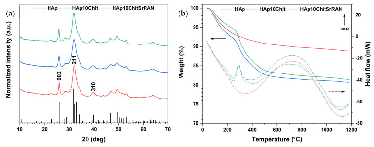
In the biomedical field, nanocrystalline hydroxyapatite is still one of the most attractive candidates as a bone substitute material due to its analogies with native bone mineral features regarding chemical composition, bioactivity and osteoconductivity. Ion substitution and low crystallinity are also fundamental characteristics of bone apatite, making it metastable, bioresorbable and reactive. In the present work, biomimetic apatite and apatite/chitosan composites were produced by dissolution-precipitation synthesis, using mussel shells as a calcium biogenic source. With an eye on possible bone reconstruction and drug delivery applications, apatite/chitosan composites were loaded with strontium ranelate, an antiosteoporotic drug. Due to the metastability and temperature sensitivity of the produced composites, sintering could be carried out by conventional methods, and therefore, cold sintering was selected for the densification of the materials. The composites were consolidated up to ~90% relative density by applying a uniaxial pressure up to 1.5 GPa at room temperature for 10 min. Both the synthesised powders and cold-sintered samples were characterised from a physical and chemical point of view to demonstrate the effective production of biomimetic apatite/chitosan composites from mussel shells and exclude possible structural changes after sintering. Preliminary in vitro tests were also performed, which revealed a sustained release of strontium ranelate for about 19 days and no cytotoxicity towards human osteoblastic-like cells (MG63) exposed up to 72 h to the drug-containing composite extract.

Keywords: apatite/chitosan composites; chitosan; cold sintering; dissolution–precipitation synthesis; drug delivery; mussel shells; nanocrystalline apatite; strontium ranelate.


Conflict of interest statement

The authors declare no conflicts of interest.

Figures

Figure 1
Figure 1
Dissolution–precipitation synthesis of SrRAN-loaded HAp/chitosan composite. The figure was created with Biorender.com.
Figure 2
Figure 2
(a) XRD patterns of HAp and HAp-based composite powders. The HAp reference pattern corresponds to No. 00-064-0738 in the ICDD database; (b) TGA-DSC curves of HAp and HAp-based composite powders. The solid lines represent the weight % of the TGA curves, while the dotted lines represent the heat flow (in mW) of the DSC curves.
Figure 3
Figure 3
SEM images of HAp (a,d), HAp10Chit (b,e) and HAp10ChitSrRAN (c,f) powder.
Figure 4
Figure 4
(a) FTIR and (b) Raman spectra of HAp, HAp10Chit and HAp10ChitSrRAN powders; (c) Raman maps indicating the distribution of apatite (green) corresponding to the ν1PO4 peak at 962 cm−1, shell organics (red) corresponding to the N–H peak at 1385 cm−1, chitosan (red) corresponding to the C–H peaks at 2929 cm−1 and SrRAN (blue) corresponding to the C≡N peak at 2204 cm−1.
Figure 5
Figure 5
Decomposition of carbonates (a) and phosphates (b) band domains of the FTIR spectra of HAp, HAp10Chit and HAp10ChitSrRAN synthesised powders.
Figure 6
Figure 6
Relative area of peaks resulting from the decomposition of ν4PO4 (a) and ν2CO3 (b) domains of HAp (red), HAp10Chit (blue) and HAp10ChitSrRAN (green) powder. (Ap. = apatitic; Non-Ap. = non apatitic).
Figure 7
Figure 7
Relative apparent density of cold-sintered HAp, HAp10Chit and HAp10ChitSrRAN. The cold-sintered pellets were produced at room temperature by holding the applied pressure for 10 min.
Figure 8
Figure 8
XRD patterns of HAp (a) and HAp10ChitSrRAN (b) pellets cold-sintered at room temperature under pressure from 250 MPa to 1500 MPa and 10 min holding time. The black triangles indicate the main peaks undergoing modifications after cold sintering.
Figure 9
Figure 9
(a) FTIR spectra of cold-sintered pellets (plt) at 1500 MPa in comparison with raw powders (pdr); (b) FTIR spectra of cold-sintered HAp10ChitSrRAN pellets pressed at 250 MPa, 500 MPa, 1000 MPa and 1500 MPa.
Figure 10
Figure 10
Cytotoxicity assessment of HAp, HAp10Chit and HAp10ChitSrRAN cold-sintered pellets by indirect test according to the ISO 10993-5:2009 standard: survival rate at 24 h, 48 h and 72 h of MG63 cells in contact with various volume of extract (1, 10 or 100 μL). The black dotted line represents the viability threshold. (*: p-value < 0.05, **: p-value < 0.005, ***: p-value < 0.001).
Figure 11
Figure 11
SrRAN release from cold-sintered HAp10ChitSrRAN pellets in DMEM and PBS expressed in % (a) and g/mL (b). The orange curve represents the release in DMEM, and the green line represents the release in PBS. (*: p-value < 0.05, ***: p-value < 0.005, ****: p-value < 0.0001).

References

    1. Wang G.C., Lu Z.F., Zreiqat H. Bone Substitute Biomaterials. Elsevier; Amsterdam, The Netherlands: 2014. Bioceramics for skeletal bone regeneration; pp. 180–216.
    1. Dorozhkin S.V. Current State of Bioceramics. J. Ceram. Sci. Technol. 2018;9:353–370. doi: 10.4416/JCST2018-00026. - DOI
    1. Eliaz N., Metoki N. Calcium Phosphate Bioceramics: A Review of Their History, Structure, Properties, Coating Technologies and Biomedical Applications. Materials. 2017;10:334. doi: 10.3390/ma10040334. - DOI - PMC - PubMed
    1. Fiume E., Magnaterra G., Rahdar A., Verné E., Baino F. Hydroxyapatite for Biomedical Applications: A Short Overview. Ceramics. 2021;4:542–563. doi: 10.3390/ceramics4040039. - DOI
    1. Bose S., Roy M., Bandyopadhyay A. Recent advances in bone tissue engineering scaffolds. Trends Biotechnol. 2012;30:546–554. doi: 10.1016/j.tibtech.2012.07.005. - DOI - PMC - PubMed

LinkOut - more resources