. 2024 Mar 2;14(5):462.
doi: 10.3390/nano14050462.

Tailoring Mesoporous Silica-Coated Silver Nanoparticles and Polyurethane-Doped Films for Enhanced Antimicrobial Applications

Affiliations

Tailoring Mesoporous Silica-Coated Silver Nanoparticles and Polyurethane-Doped Films for Enhanced Antimicrobial Applications

Silvia Nuti et al. Nanomaterials (Basel). .

Abstract

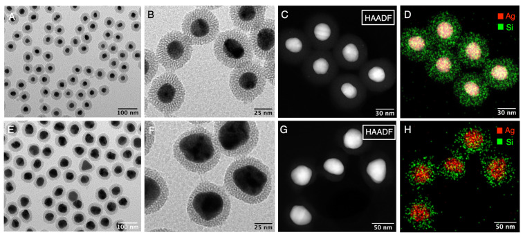
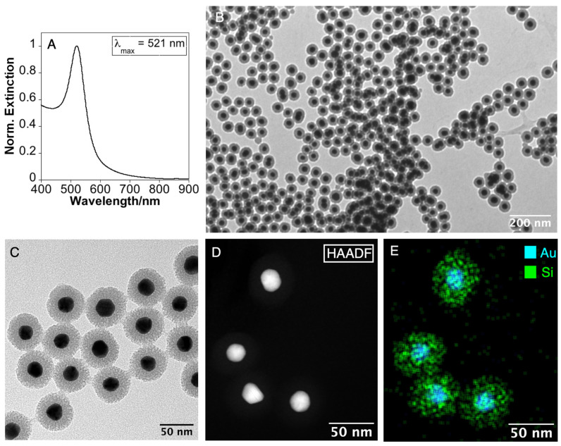
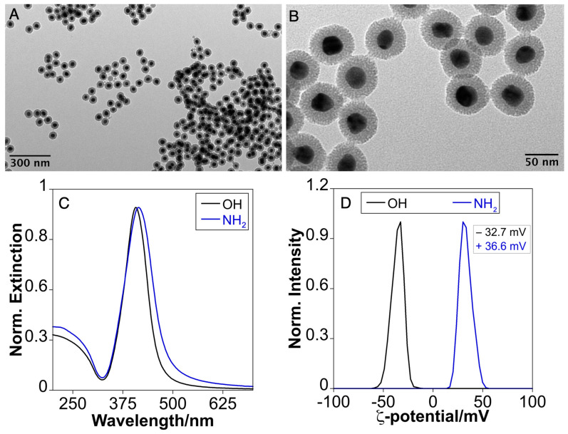
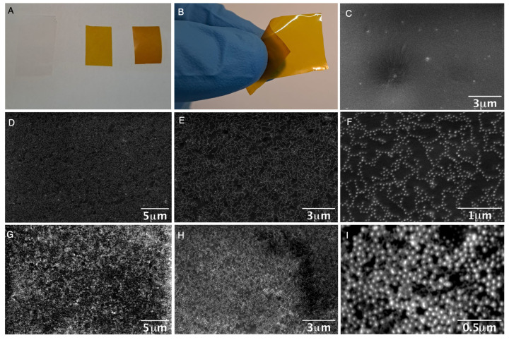
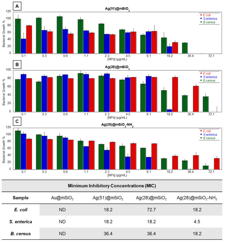
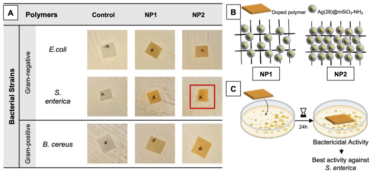
The global increase in multidrug-resistant bacteria poses a challenge to public health and requires the development of new antibacterial materials. In this study, we examined the bactericidal properties of mesoporous silica-coated silver nanoparticles, varying the core sizes (ca. 28 nm and 51 nm). We also investigated gold nanoparticles (ca. 26 nm) coated with mesoporous silica as possible inert metal cores. To investigate the modification of antimicrobial activity after the surface charge change, we used silver nanoparticles with a silver core of 28 nm coated with a mesoporous shell (ca. 16 nm) and functionalized with a terminal amine group. Furthermore, we developed a facile method to create mesoporous silica-coated silver nanoparticles (Ag@mSiO2) doped films using polyurethane (IROGRAN®) as a polymer matrix via solution casting. The antibacterial effects of silver nanoparticles with different core sizes were analyzed against Gram-negative and Gram-positive bacteria relevant to the healthcare and food industry. The results demonstrated that gold nanoparticles were inert, while silver nanoparticles exhibited antibacterial effects against Gram-negative (Escherichia coli and Salmonella enterica subsp. enterica serovar Choleraesuis) and Gram-positive (Bacillus cereus) strains. In particular, the larger Ag@mSiO2 nanoparticles showed a minimum inhibitory concentration (MIC) and a minimum bactericidal concentration (MBC) of 18 µg/mL in the Salmonella strain. Furthermore, upon terminal amine functionalization, reversing the surface charge to positive values, there was a significant increase in the antibacterial activity of the NPs compared to their negative counterparts. Finally, the antimicrobial properties of the nanoparticle-doped polyurethane films revealed a substantial improvement in antibacterial efficacy. This study provides valuable information on the potential of mesoporous silica-coated silver nanoparticles and their applications in fighting multidrug-resistant bacteria, especially in the healthcare and food industries.

Keywords: antimicrobial activity; bacterials; gold nanoparticles; polymers; silica core shell; silver nanoparticles.


Conflict of interest statement

The authors declare no conflicts of interest.

Figures

Figure 1
Figure 1
(A,B) Low-magnification TEM images of Ag(28)@mSiO2. (C) High-magnification HAADF-STEM image and (D) Ag and Si distribution EDX maps of Ag(28)@mSiO2. (E,F) Low-magnification TEM images of Ag(51)@mSiO2. (C,G) High-magnification HAADF-STEM image and (D,H) Ag and Si distribution EDX maps of Ag(28)@mSiO2 and Ag(51)@mSiO2 respectively.
Figure 2
Figure 2
(A) UV-Vis extinction spectrum of Au NPs. (B,C) Low-magnification TEM images of Au@mSiO2. (D) High-magnification HAADF-STEM image and (E) Au and Si distribution EDX maps of Au@mSiO2 NPs.
Figure 3
Figure 3
(A,B) Low-magnification TEM images of Ag(28)@mSiO2-NH2, (C) UV-Vis extinction spectrum and (D) ζ-potential of purified Ag(28)@mSiO2-OH (black) and Ag(28)@mSiO2-NH2 (blue).
Figure 4
Figure 4
(A) Undoped film (transparent film) and doped film with different quantity of Ag(28)@mSiO2-NH2 nanoparticles (yellow films). (B) Flexibility of the doped film. (C,D) Scanning Electron Microscope (SEM) top-view images of the undoped film (C) and doped film with 4 μg/cm2 (DF) and 8 μg/cm2 (GI) at different magnifications (10K×: (D,G); 20K×: (C,E,H); 80K×: (F) and 160K×: (I)).
Figure 5
Figure 5
(A) SEM images of the cross-section view of the undoped polymer and doped film with 4 μg/cm2 (B) and 8 μg/cm2 (C). SEM images at different magnification of different areas of the cross section where the nanoparticles are exposed towards the surface for the film doped with 4 μg/cm2 (D,E) and 8 μg/cm2 (F,G).
Figure 6
Figure 6
(AC) Bacterial growth profiles, against Gram-negative bacteria (Red) Escherichia coli (E. coli, ATCC® 25922TM), (Blue) Salmonella enterica subsp. enterica serovar Choleraesuis (ATCC® 10708TM) and Gram-positive bacteria (Green) Bacillus cereus (ATCC® 11778TM). Concentrations are relative to the metal concentration in the nanoparticles previously determined via ICP. Obtained MICs for all nanoparticles against the tested Gram-negative and Gram-positive bacteria. In the case of S. enterica the obtained MICs correspond to the MBC values, as well. NPs: Au@mSiO2; Ag(51)@mSiO2; Ag(28)@mSiO2; Ag(28)@mSiO2-NH2.
Figure 7
Figure 7
Assessment of antibacterial activity of Ag(28)@mSiO2-NH2-doped IROGRAN polymers (A). Polymers were doped with a lower (NP1) and higher (NP2) NP concentration, respectively. Bacterial strains selected were Escherichia coli (E. coli, ATCC® 25922TM), Salmonella enterica subsp. enterica serovar Choleraesuis (ATCC® 10708TM) and Bacillus cereus (ATCC® 11778TM). Areas of all polymeric squares ca. 1 cm2. Red highlight indicates the best obtained results, through the occurrence of a slight inhibitory halo. Schematic overview of the different polymeric matrixes applied (B) and the assay conducted (C).

Similar articles

Cited by

References

    1. Brown E.D., Wright G.D. Antibacterial Drug Discovery in the Resistance Era. Nature. 2016;529:336–343. doi: 10.1038/nature17042. - DOI - PubMed
    1. Kaper J.B., Nataro J.P., Mobley H.L.T. Pathogenic Escherichia coli. Nat. Rev. Microbiol. 2004;2:123–140. doi: 10.1038/nrmicro818. - DOI - PubMed
    1. Blair J.M.A., Webber M.A., Baylay A.J., Ogbolu D.O., Piddock L.J.V. Molecular Mechanisms of Antibiotic Resistance. Nat. Rev. Microbiol. 2015;13:42–51. doi: 10.1038/nrmicro3380. - DOI - PubMed
    1. Wales A.D., Allen V.M., Davies R.H. Chemical Treatment of Animal Feed and Water for the Control of Salmonella. Foodborne Pathog. Dis. 2010;7:3–15. doi: 10.1089/fpd.2009.0373. - DOI - PubMed
    1. Nowack B., Krug H.F., Height M. 120 Years of Nanosilver History: Implications for Policy Makers. Environ. Sci. Technol. 2011;45:7593–7595. doi: 10.1021/es2017895. - DOI - PubMed

LinkOut - more resources