Review
. 2024 Mar 4;14(5):465.
doi: 10.3390/nano14050465.

Recent Advances in Tactile Sensory Systems: Mechanisms, Fabrication, and Applications

Affiliations
Review

Recent Advances in Tactile Sensory Systems: Mechanisms, Fabrication, and Applications

Jianguo Xi et al. Nanomaterials (Basel). .

Abstract

Flexible electronics is a cutting-edge field that has paved the way for artificial tactile systems that mimic biological functions of sensing mechanical stimuli. These systems have an immense potential to enhance human-machine interactions (HMIs). However, tactile sensing still faces formidable challenges in delivering precise and nuanced feedback, such as achieving a high sensitivity to emulate human touch, coping with environmental variability, and devising algorithms that can effectively interpret tactile data for meaningful interactions in diverse contexts. In this review, we summarize the recent advances of tactile sensory systems, such as piezoresistive, capacitive, piezoelectric, and triboelectric tactile sensors. We also review the state-of-the-art fabrication techniques for artificial tactile sensors. Next, we focus on the potential applications of HMIs, such as intelligent robotics, wearable devices, prosthetics, and medical healthcare. Finally, we conclude with the challenges and future development trends of tactile sensors.

Keywords: HMIs; fabrication techniques; mechanism; robotics; tactile sensors.


Conflict of interest statement

The authors declare no conflicts of interest.

Figures
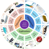
Figure 1
Figure 1
Schematic illustration of tactile sensors from mechanisms, fabrications, and applications. (a) Reproduced with permission from Ref. [34]. Copyright 2023, Springer Nature. (b) Reproduced with permission from Ref. [35]. Copyright 2018, Springer Nature. (c) Reproduced with permission from Ref. [36]. Copyright 2022, American Chemistry Society. (d) Reproduced with permission from Ref. [37]. Copyright 2021, Wiley. (e) Reproduced with permission from Ref. [38]. Copyright 2021, Science. (f) Reproduced with permission from Ref. [39]. Copyright 2023, Spinger Nature. (g) Reproduced with permission from Ref. [40]. Copyright 2022, Spinger Nature. (h) Reproduced with permission from Ref. [41]. Copyright 2020, Spinger Nature. (i) Reproduced with permission from Ref. [42]. Copyright 2023, Spinger Nature. (j) Reproduced with permission from Ref. [43]. Copyright 2021, Elsevier. (k) Reproduced with permission from Ref. [44]. Copyright 2023, Springer Nature. (l) Reproduced with permission from Ref. [45]. Copyright 2011, Springer Nature.
Figure 2
Figure 2
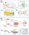
(a) The basic procedure of the synaptic current activated by external stimuli in the biological tactile system. (b) Schematic illustration of four typical working principles of flexible tactile sensor, (i) piezoresistive, (ii) capacitive, (iii) piezoelectric, and (iv) triboelectric.
Figure 3
Figure 3
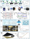
(a) Sectional view of porous AgNWs/PVDF composite (i) with the sensitivity under various pressure ranges (ii). Reproduced with permission from Ref. [46]. Copyright 2022, American Chemical Society. (b) SEM image of the porous PDMS with CNTs/graphene layer (i). (ii) Sensitivities of the flexible pressure sensor when coating by CNTs/graphene, CNTs, and graphene layers. Reproduced with permission from Ref. [47]. Copyright 2022, American Chemical Society. (c) SEM images of PDMS/CNT microstructures (ii) molded by the stainless-steel mesh (i), which was treated as a frame for PDMS/CNT microstructure. Reproduced with permission from Ref. [48]. Copyright 2020, Wiley. (d) Schematic illustration of the soft robotic earthworm, which was moving through a cylinder array (i). (ii) Photograph of serpentine piezoresistive strain sensors. Reproduced with permission from Ref. [49]. Copyright 2020, American Chemical Society.
Figure 4
Figure 4
Schematic illustration of the fabrication of neuromorphic tactile systems. (a) The NeuTap concept design. (i) The fusion of spatial and temporal pattern recognition within the tactile perception-feedback loop. (ii) The structure of a sensory neuron’s function. (iii) The illustrative details for the intricate working process and components of the NeuTap system. Reproduced with permission from Ref. [55]. Copyright 2018, Wiley. (b) The structure of an artificial afferent nerve system involves pressure sensors, an organic ring oscillator, and a synaptic transistor for signal transduction, in which the single ring oscillator is connected to a synaptic transistor. The architecture allows for the integration of multiple ring oscillators, each linked to a cluster of pressure sensors, converging on a solitary synaptic transistor. Reproduced with permission from Ref. [56]. Copyright 2018, Science.
Figure 5
Figure 5
Structural illustration of capacitive tactile sensors. (a) A flexible pressure sensor based on μ-graphene electrodes. (i) The structure of MGr-based pressure sensor. (ii) Sensing mechanism of MGr-based pressure sensor. (iii) The response time of MGr-based pressure sensor. Reproduced with permission from Ref. [59]. Copyright 2019, American Chemical Society. (b) The structure design of the DOT-TPS (i). (ii) Equivalent circuit of the DOT-TPS. (iii) The relative changes of Ipost and the response time for DOT-TPS when applying different pressures. Reproduced with permission from Ref. [63]. Copyright 2017, Wiley.
Figure 6
Figure 6
(a) The human sensory nervous system features mechanoreceptors in the skin that transform mechanical stimuli into presynaptic potential signals, which are then transmitted to the central nervous system via neurons and synapses (i). (ii) Schematic diagram of the graphene artificial sensory synapse based on piezotronics effect. (iii) An equivalent circuit diagram of the piezoelectric potential’s interaction through an ion gel. Reproduced with permission from Ref. [70]. Copyright 2019, Wiley. (b) In the human reflex arc, stimulation of a thigh muscle induces a response where the sensory neuron communicates an action potential to the spinal cord’s gray matter. Within the spinal cord, this neuron forms a direct synaptic link with a motor neuron. If the stimulus is sufficiently intense, it can initiate action potentials in the motor neuron, resulting in a knee jerk (i). (ii) The Strain-Powered Device (SPD) uses an external strain on a cantilever mechanism to mimic the logical processing of a human reflex, modulating output power based on mechanical input. Reproduced with permission from Ref. [72]. Copyright 2020, Springer Nature.
Figure 7
Figure 7
(a) Schematic illustration of triboelectric tactile systems activated by contact electrification, which includes a self-activation component, a synaptic transistor, and a functional circuit. (b) Schematic diagram of the CE-activated MoS2 synaptic transistor with annotations of each component. (c) Equivalent circuit of the CE-activated artificial afferents. Reproduced with permission from Ref. [84]. Copyright 2021, Springer Nature.
Figure 8
Figure 8
(a) Schematic illustration of bimodal sensor fabricated by inkjet printing (i). (ii) The durability of the strain sensor at a strain of 0.76% and 1.07%, respectively. (iii) The durability of the pressure sensor at a pressure of 202.8 kPa. Reproduced with permission from Ref. [96]. Copyright 2019, Wiley. (b) Schematic illustration of the fabrication process of PUA/Ag composite film as flexible electrode (i). (ii) Durability under 1% strain with a frequency of 0.1 Hz and 1000 stretching/releasing cycles. (iii) The response time of single-layer PUA/Ag-based strain sensor under 1% strain. Reproduced with permission from Ref. [97]. Copyright 2021, Wiley.
Figure 9
Figure 9
(a) Flow chart of the fabrication of e-skin 3D printing based on a porous ionic gel (i) with a high sensitivity of 0.213 kPa−1. (ii) Pressure vs resistance response of the e-skin pressure sensor. Reproduced with permission from Ref. [100]. Copyright 2022, American Chemistry Society. (b) Surface-embedded graphene sensors. (i) SEG sensors are fabricated in the following procedures: An ABS mold was made via 3D printing and then was dip coating in the graphene solution repeatedly. After dehydration by hot air flow, graphene nanoplatelets (GnP) was adhered to the surface of ABS mold uniformly. Subsequently, silicone rubber (SR) was cast, leading to GnP transfer to the SR surface after dissolving ABS mold with resistance changes (ii) and stable gauge factors (iii) when applying 200 cycles and 0~400 cycles under strain of 2%, 5%, 10%. Reproduced with permission from Ref. [101]. Copyright 2020, American Chemistry Society. (c) Schematic illustration of DLP 3D printing for wearable devices with different structures (i). (ii) The image of the compression/release state of the lattice-structure sensor under 85% strain. (iii) The pressure sensitivity when sensors are lattice-structured or bulk-structured. Reproduced with permission from Ref. [102]. Copyright 2022, Elsevier. (d) The structure design of the sandpaper morphology and PDMS/CNTs (SPC) sensor consisting of the SPC film and interdigitated electrode layers (i). (ii) The sensitivity of the 600-mesh-roughness SPC sensor within the range of 10 kPa. Reproduced with permission from Ref. [103]. Copyright 2022, American Chemistry Society.
Figure 10
Figure 10
(a) Schematic illustration of a robotic tactile system, in which pressure stimuli applied to mechanoreceptors induce changes in the receptor potential and then initiate action potentials. Action potentials activated by multiple nerve fibers are superposed through neurons and used for information processing, resulting in the synaptic network of the brain recognizing the input pressure pattern. Reproduced with permission from Ref. [1]. Copyright 2020, Springer Nature. (b) Schematic illustration of gesture recognition and control of the tactile sensor system (i). (ii) The structural design of the tactile sensor. (iii) SEM image of PVDF nanowires prepared via electrospinning with a scale bar of 5 μm. (iv) XRD pattern of PVDF film. Reproduced with permission from Ref. [143]. Copyright 2022, American Chemistry Society. (c) The structural diagram of the multilayered e-dermis over the fingertips of a prosthetic hand (i), including conductive and piezoresistive textiles encased in rubber, in which a dermal layer of two piezoresistive sensing elements is separated from the epidermal layer with a silicone rubber layer of 1 mm. (ii) The counterparts of humans’ biological mechanoreceptors in healthy glabrous skin. Reproduced with permission from Ref. [144]. Copyright 2018, Science. (d) The procedures that a biological system uses to perceive (i) and process (ii) external pressure. Schematic diagram of the volatile pristine in-memory tactile sensor (iii) and the nonvolatile PEI de-doped in-memory tactile sensor (iv). Reproduced with permission from Ref. [145]. Copyright 2023, American Chemistry Society.

Similar articles

Cited by

References

    1. Kim S., Lee Y., Kim H.-D., Choi S.-J. A tactile sensor system with sensory neurons and a perceptual synaptic network based on semivolatile carbon nanotube transistors. NPG Asia Mater. 2020;12:76. doi: 10.1038/s41427-020-00258-9. - DOI
    1. Skedung L., Arvidsson M., Chung J.Y., Stafford C.M., Berglund B., Rutland M.W. Feeling Small: Exploring the Tactile Perception Limits. Sci. Rep. 2013;3:2617. doi: 10.1038/srep02617. - DOI - PMC - PubMed
    1. Chun S., Kim J.-S., Yoo Y., Choi Y., Jung S.J., Jang D., Lee G., Song K.-I., Nam K.S., Youn I., et al. An artificial neural tactile sensing system. Nat. Electron. 2021;4:429–438. doi: 10.1038/s41928-021-00585-x. - DOI
    1. Luo Y., Li Y., Sharma P., Shou W., Wu K., Foshey M., Li B., Palacios T., Torralba A., Matusik W. Learning human–environment interactions using conformal tactile textiles. Nat. Electron. 2021;4:193–201. doi: 10.1038/s41928-021-00558-0. - DOI
    1. Hu Z., Lin L., Lin W., Xu Y., Xia X., Peng Z., Sun Z., Wang Z. Machine Learning for Tactile Perception: Advancements, Challenges, and Opportunities. Adv. Intell. Syst. 2023;5:2200371. doi: 10.1002/aisy.202200371. - DOI

LinkOut - more resources