

Multi-modal deep learning methods for classification of chest diseases using different medical imaging and cough sounds
- PMID: 38470893
- PMCID: PMC10931489
- DOI: 10.1371/journal.pone.0296352
Multi-modal deep learning methods for classification of chest diseases using different medical imaging and cough sounds
Abstract
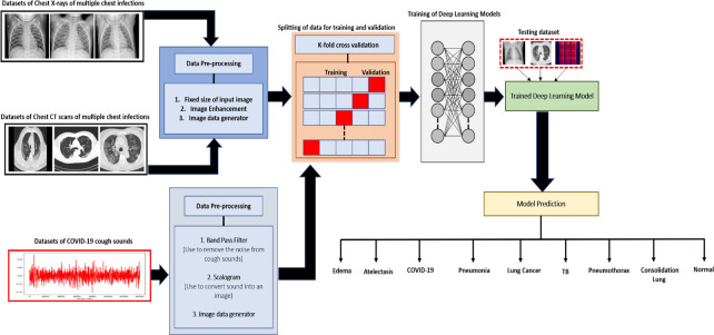
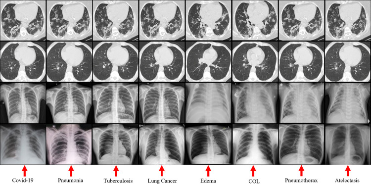
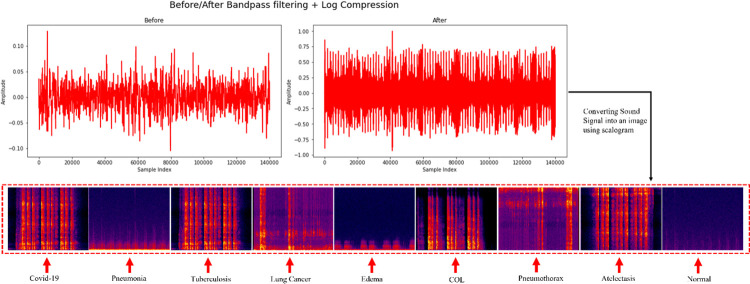
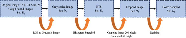
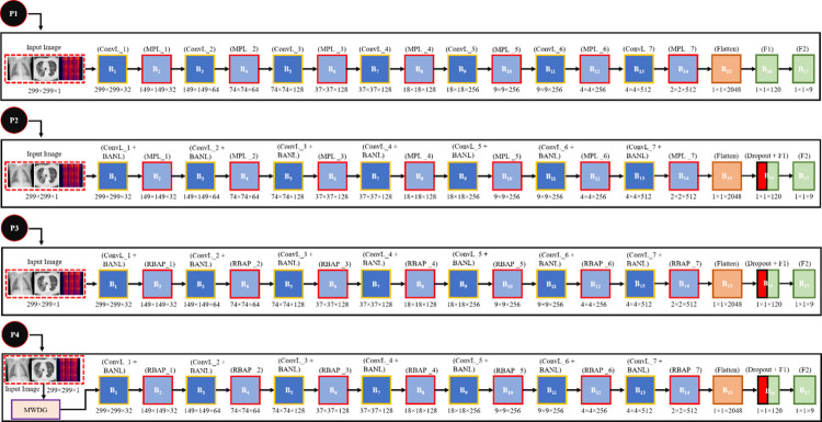
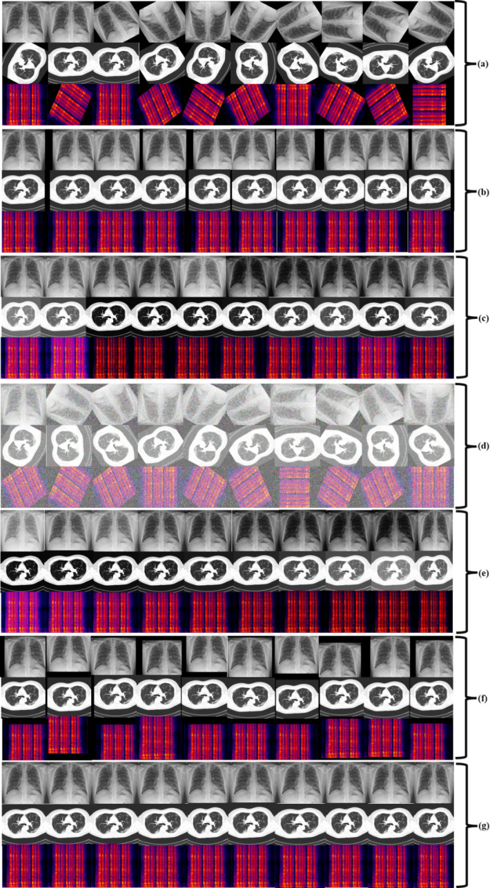
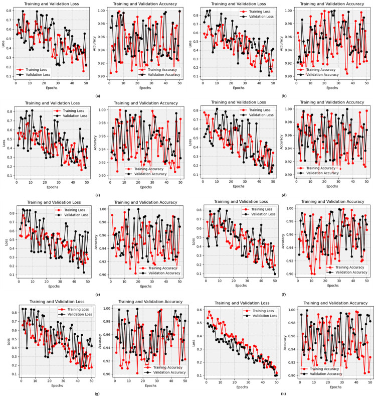
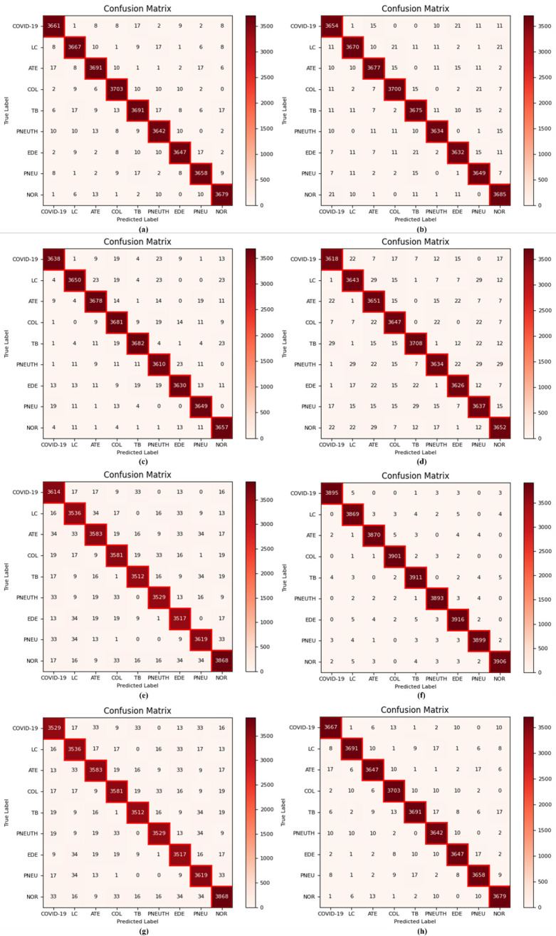
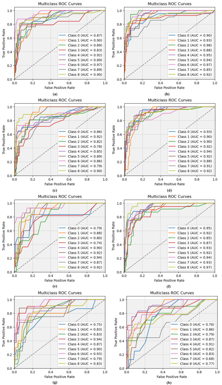
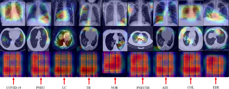
Chest disease refers to a wide range of conditions affecting the lungs, such as COVID-19, lung cancer (LC), consolidation lung (COL), and many more. When diagnosing chest disorders medical professionals may be thrown off by the overlapping symptoms (such as fever, cough, sore throat, etc.). Additionally, researchers and medical professionals make use of chest X-rays (CXR), cough sounds, and computed tomography (CT) scans to diagnose chest disorders. The present study aims to classify the nine different conditions of chest disorders, including COVID-19, LC, COL, atelectasis (ATE), tuberculosis (TB), pneumothorax (PNEUTH), edema (EDE), pneumonia (PNEU). Thus, we suggested four novel convolutional neural network (CNN) models that train distinct image-level representations for nine different chest disease classifications by extracting features from images. Furthermore, the proposed CNN employed several new approaches such as a max-pooling layer, batch normalization layers (BANL), dropout, rank-based average pooling (RBAP), and multiple-way data generation (MWDG). The scalogram method is utilized to transform the sounds of coughing into a visual representation. Before beginning to train the model that has been developed, the SMOTE approach is used to calibrate the CXR and CT scans as well as the cough sound images (CSI) of nine different chest disorders. The CXR, CT scan, and CSI used for training and evaluating the proposed model come from 24 publicly available benchmark chest illness datasets. The classification performance of the proposed model is compared with that of seven baseline models, namely Vgg-19, ResNet-101, ResNet-50, DenseNet-121, EfficientNetB0, DenseNet-201, and Inception-V3, in addition to state-of-the-art (SOTA) classifiers. The effectiveness of the proposed model is further demonstrated by the results of the ablation experiments. The proposed model was successful in achieving an accuracy of 99.01%, making it superior to both the baseline models and the SOTA classifiers. As a result, the proposed approach is capable of offering significant support to radiologists and other medical professionals.
Copyright: © 2024 Malik, Anees. This is an open access article distributed under the terms of the Creative Commons Attribution License, which permits unrestricted use, distribution, and reproduction in any medium, provided the original author and source are credited.
Conflict of interest statement
The authors have declared that no competing interests exist.
Figures
Similar articles
-
Deep Learning-Based Classification of Chest Diseases Using X-rays, CT Scans, and Cough Sound Images.Diagnostics (Basel). 2023 Aug 26;13(17):2772. doi: 10.3390/diagnostics13172772. Diagnostics (Basel). 2023. PMID: 37685310 Free PMC article.
-
CDC_Net: multi-classification convolutional neural network model for detection of COVID-19, pneumothorax, pneumonia, lung Cancer, and tuberculosis using chest X-rays.Multimed Tools Appl. 2023;82(9):13855-13880. doi: 10.1007/s11042-022-13843-7. Epub 2022 Sep 20. Multimed Tools Appl. 2023. PMID: 36157356 Free PMC article.
-
DMFL_Net: A Federated Learning-Based Framework for the Classification of COVID-19 from Multiple Chest Diseases Using X-rays.Sensors (Basel). 2023 Jan 9;23(2):743. doi: 10.3390/s23020743. Sensors (Basel). 2023. PMID: 36679541 Free PMC article.
-
Machine and Deep Learning for Tuberculosis Detection on Chest X-Rays: Systematic Literature Review.J Med Internet Res. 2023 Jul 3;25:e43154. doi: 10.2196/43154. J Med Internet Res. 2023. PMID: 37399055 Free PMC article.
-
Development and integration of VGG and dense transfer-learning systems supported with diverse lung images for discovery of the Coronavirus identity.Inform Med Unlocked. 2022;32:101004. doi: 10.1016/j.imu.2022.101004. Epub 2022 Jul 8. Inform Med Unlocked. 2022. PMID: 35822170 Free PMC article. Review.
Cited by
-
LGD_Net: Capsule network with extreme learning machine for classification of lung diseases using CT scans.PLoS One. 2025 Aug 8;20(8):e0327419. doi: 10.1371/journal.pone.0327419. eCollection 2025. PLoS One. 2025. PMID: 40779565 Free PMC article.
-
Improved swin transformer-based thorax disease classification with optimal feature selection using chest X-ray.PLoS One. 2025 Jun 25;20(6):e0327099. doi: 10.1371/journal.pone.0327099. eCollection 2025. PLoS One. 2025. PMID: 40561187 Free PMC article.
References
-
- WHO Coronavirus (COVID-19) Dashboard|WHO Coronavirus (COVID-19) Dashboard with Vaccination Data. Available online: https://covid19.who.int/ (accessed on 10th February 2023).
-
- Arevalo-Rodriguez Ingrid, Diana Buitrago-Garcia Daniel Simancas-Racines, Paula Zambrano-Achig Rosa Del Campo, Ciapponi Agustin, Sued Omar et al.. "False-negative results of initial RT-PCR assays for COVID-19: a systematic review." PloS one 15, no. 12 (2020): e0242958. doi: 10.1371/journal.pone.0242958 - DOI - PMC - PubMed
MeSH terms
LinkOut - more resources
Full Text Sources
Medical
Research Materials
