Review
. 2024 Apr 2;57(7):1019-1031.
doi: 10.1021/acs.accounts.3c00759. Epub 2024 Mar 12.

FSHing for DNA Damage: Key Features of MutY Detection of 8-Oxoguanine:Adenine Mismatches

Affiliations
Review

FSHing for DNA Damage: Key Features of MutY Detection of 8-Oxoguanine:Adenine Mismatches

Chandrima Majumdar et al. Acc Chem Res. .

Abstract

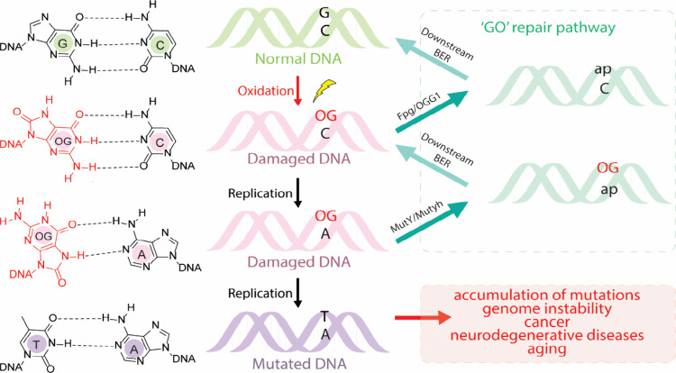
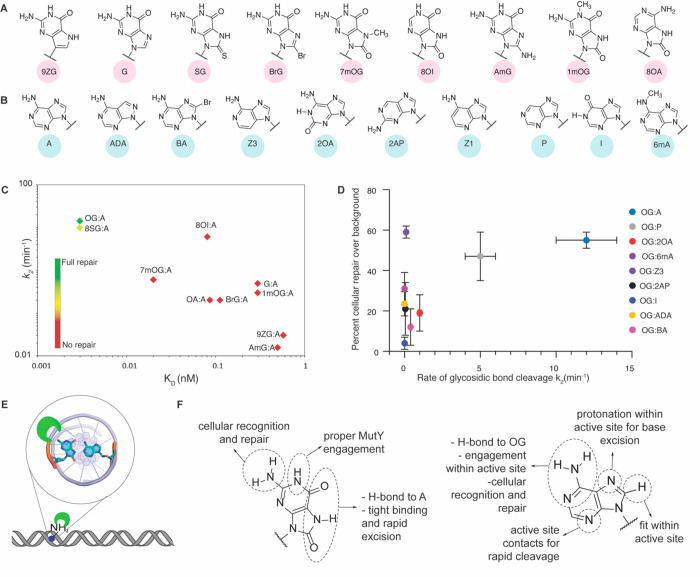
Base excision repair (BER) enzymes are genomic superheroes that stealthily and accurately identify and remove chemically modified DNA bases. DNA base modifications erode the informational content of DNA and underlie many disease phenotypes, most conspicuously, cancer. The "OG" of oxidative base damage, 8-oxo-7,8-dihydroguanine (OG), is particularly insidious due to its miscoding ability that leads to the formation of rare, pro-mutagenic OG:A mismatches. Thwarting mutagenesis relies on the capture of OG:A mismatches prior to DNA replication and removal of the mis-inserted adenine by MutY glycosylases to initiate BER. The threat of OG and the importance of its repair are underscored by the association between inherited dysfunctional variants of the MutY human homologue (MUTYH) and colorectal cancer, known as MUTYH-associated polyposis (MAP). Our functional studies of the two founder MUTYH variants revealed that both have compromised activity and a reduced affinity for OG:A mismatches. Indeed, these studies underscored the challenge of the recognition of OG:A mismatches that are only subtly structurally different than T:A base pairs. Since the original discovery of MAP, many MUTYH variants have been reported, with most considered to be "variants of uncertain significance." To reveal features associated with damage recognition and adenine excision by MutY and MUTYH, we have developed a multipronged chemical biology approach combining enzyme kinetics, X-ray crystallography, single-molecule visualization, and cellular repair assays. In this review, we highlight recent work in our laboratory where we defined MutY structure-activity relationship (SAR) studies using synthetic analogs of OG and A in cellular and in vitro assays. Our studies revealed the 2-amino group of OG as the key distinguishing feature of OG:A mismatches. Indeed, the unique position of the 2-amino group in the major groove of OGsyn:Aanti mismatches provides a means for its rapid detection among a large excess of highly abundant and structurally similar canonical base pairs. Furthermore, site-directed mutagenesis and structural analysis showed that a conserved C-terminal domain β-hairpin "FSH'' loop is critical for OG recognition with the "His" serving as the lesion detector. Notably, MUTYH variants located within and near the FSH loop have been associated with different forms of cancer. Uncovering the role(s) of this loop in lesion recognition provided a detailed understanding of the search and repair process of MutY. Such insights are also useful to identify mutational hotspots and pathogenic variants, which may improve the ability of physicians to diagnose the likelihood of disease onset and prognosis. The critical importance of the "FSH" loop in lesion detection suggests that it may serve as a unique locus for targeting probes or inhibitors of MutY/MUTYH to provide new chemical biology tools and avenues for therapeutic development.


Conflict of interest statement

The authors declare no competing financial interest.

Figures

Figure 1
Figure 1
Presence of OG in DNA leads to the formation of G:C to T:A transversion mutations. The GO repair pathway features two base excision repair (BER) glycosylases, Fpg/OGG1 and MutY/MUTYH, that prevent mutations associated with OG by removing OG from OG:C base pairs or preventing propagation of the pro-mutagenic OG:A base pairs by removal of the misplaced A, respectively. The abasic site (designated as “ap”) product is removed and replaced using the intact strand as the guide by downstream BER pathway enzymes. Note that replicative polymerases more frequently insert “A” over “C” opposite OG while repair polymerases (β/λ) exhibit a higher tendency to incorporate “C” over “A” opposite OG.
Figure 2
Figure 2
Crystal structures of Gs MutY reveal details of the catalytic mechanism. (A) Overall structure of Gs MutY bound to DNA containing transition-state analog 1N across OG (PDB ID 6U7T). (B) Extruded 1N in the active site and active site residues (PDB ID 6U7T). (C) Key residues from the C- and N-terminal domain form a network of interactions around OGanti. (D) Contacts observed in FLRC (PDB ID 3G0Q) to the A base flipped into the active site within the N-terminal domain. (E) Proposed mechanism for MutY-mediated adenine excision. Protonation of the A at N7 enables it to leave as a neutral molecule. The oxacarbenium ion hence formed is stabilized by the formation of a transient acetal covalent intermediate with the catalytic aspartate; subsequent hydrolysis of the acetal intermediate leads to the formation of the abasic site product. Panel E was adapted with permission from ref (38).
Figure 3
Figure 3
Schematic description of assays used to evaluate different stages of repair. (A) Minimal kinetics scheme depicting the major steps in the enzymatic activity of MutY in terms of the binding constant, KD, rate of adenine excision, k2, and rate of abasic site product release, k3. (B) Electrophoretic mobility shift assays, using either a catalytically inactive enzyme or a nonhydrolyzable substrate, are employed to evaluate the binding affinity of the enzyme for substrate analogs. (C) Glycosylase assay used to evaluate kinetic parameters k2 and k3. (D) E. coli-based cellular repair assay used to evaluate the overall extent of repair in terms of the conversion of an OG:A mispair to G:C. An in-depth description of biochemical and cellular assay methods can be found in refs (44), (45), and (46).
Figure 4
Figure 4
Structure activity relationships (SAR) studies reveal key features needed for recognition and repair by MutY. (A) Chemical structures of OG analogs. (B) Chemical structures of adenine analogs. (C) Relationship between the rate of A cleavage and binding constants of the catalytically inactive E37S MutY variant to duplexes containing X:A mispairs, where X is the OG analog. (D) Relationship between the rate of excision of adenine analogs and overall cellular repair in E. coli. The adenine analogs exhibited tight binding (KD < 5 pM) when paired opposite OG. (E) The unique H-bonding ability of A to OG positions the 2-amino group in the major groove of DNA to enable recognition by MutY. (F) Summary of the roles of structural features of OG and A as determined by SAR analysis. Adapted with permission from refs (1) and (2), copyrights 2017 and 2020, respectively, American Chemical Society.
Figure 5
Figure 5
Recognition of OG:A by MutY is dependent on a conserved C-terminal loop. (A) FSH loop invades the DNA helix searching for OG (green, PDB ID: 6U7T). (B) Overlay of crystal structures of Gs MutY bound to the transition-state analog paired opposite OG (magenta; PDB ID: 6U7T) and G (cyan; PDB ID 6Q0C) showing the invasion of the DNA helix by the FSH loop. The FSH loop serine (S308 in Gs MutY) is proposed to aid discrimination between OG and G through the formation of an OG-specific hydrogen bond. (C) Key residues from C- and N-terminal domains form a network of interactions around OGanti while Ganti lacks the interactions (denoted with red X) with the serine residue that has rotated 120° but maintained interactions with tyrosine. (D) Graph showing the binding and catalytic specificity of FSH loop mutants. The mutation suppression frequency is represented by the size of the circles corresponding to each variant. Smaller circles indicate that the MutY variant is competent in suppressing G:C → T:A mutations in cells. Data in panel D was taken from ref (3).
Figure 6
Figure 6
Single-molecule studies using E. coli MutY revealed the role of the C-terminal domain histidine, H296, in OG:A detection. (A) Overview of single-molecule-based assay utilizing Q-Dot conjugated glycosylase molecules to monitor in real time diffusion on lesion- or nonlesion-containing stretched DNA. Lesions are equally spaced between CY5 markers to correlate periods of “pausing” with the sensing of lesions. (B) Cartoon depiction highlighting observed outcomes when WT MutY is monitored in search of OGA (top) OI:A (middle) histidine 296 searching for OG:A (bottom). (C) Representative displacement trajectories of WT MutY and H296A variant on an OG/OI:A lesion containing concatemerized substrate DNA. (D) Histogram showing counts for individual glycosylase molecules and computed diffusion rates. The figure was adapted from ref (4), copyright 2020, American Chemical Society.

References

    1. Manlove A. H.; McKibbin P. L.; Doyle E. L.; Majumdar C.; Hamm M. L.; David S. S. Structure-Activity Relationships Reveal Key Features of 8-Oxoguanine: A Mismatch Detection by the MutY Glycosylase. ACS Chem. Biol. 2017, 12, 2335–2344. 10.1021/acschembio.7b00389. - DOI - PMC - PubMed
    1. Majumdar C.; McKibbin P. L.; Krajewski A. E.; Manlove A. H.; David S. S. Unique H-Bonding Pattern of Adenine with the Oxidatively Damaged Base 8-Oxoguanine Enables Specific Recognition and Repair by DNA Glycosylase MutY. J. Am. Chem. Soc. 2020, 142, 20340–20350. 10.1021/jacs.0c06767. - DOI - PMC - PubMed
    1. Russelburg L. P.; O’Shea Murray V. L.; Demir M.; Knutsen K. R.; Sehgal S. L.; Cao S.; David S. S.; Horvath M. P. Structural Basis for Finding OG Lesions and Avoiding Undamaged G by the DNA Glycosylase MutY. ACS Chem. Biol. 2020, 15, 93–102. 10.1021/acschembio.9b00639. - DOI - PMC - PubMed
    1. Lee A. J.; Majumdar C.; Kathe S. D.; Van Ostrand R. P.; Vickery H. R.; Averill A. M.; Nelson S. R.; Manlove A. H.; McCord M. A.; David S. S. Detection of OG:A Lesion Mispairs by MutY Relies on a Single His Residue and the 2-Amino Group of 8-Oxoguanine. J. Am. Chem. Soc. 2020, 142, 13283–13287. 10.1021/jacs.0c04284. - DOI - PMC - PubMed
    1. Ziada A. S.; Smith M. S. R.; Côté H. C. F. Updating the Free Radical Theory of Aging. Front. Cell Dev. Biol. 2020, 8, 1–5. 10.3389/fcell.2020.575645. - DOI - PMC - PubMed

Publication types