. 2024 Jul 1;63(4):428-436.
doi: 10.30802/AALAS-JAALAS-23-000107. Epub 2024 Mar 12.

Determination of Postmortem Interval in Mice

Affiliations

Determination of Postmortem Interval in Mice

Rachel R Howie et al. J Am Assoc Lab Anim Sci. .

Abstract

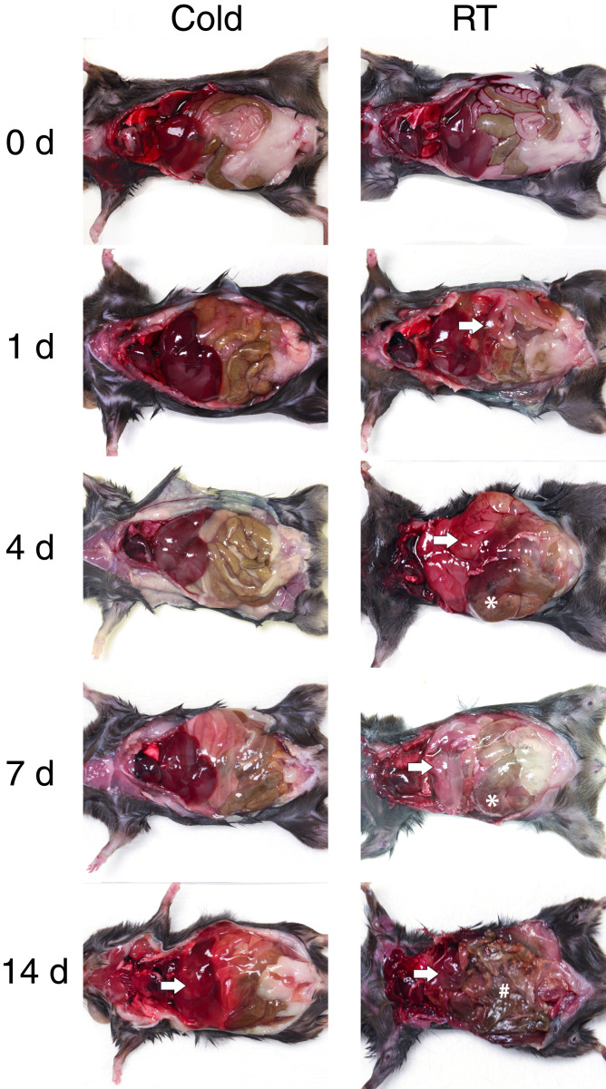
Despite the major use of mice in biomedical research, little information is available with regard to identifying their postmortem changes and using that information to determine the postmortem interval (PMI), defined as the time after death. Both PMI and environmental conditions influence decomposition (autolysis and putrefaction) and other postmortem changes. Severe decomposition compromises lesion interpretation and disease detection and wastes limited pathology resources. The goal of this study was to assess postmortem changes in mice in room temperature cage conditions and under refrigeration at 4 °C to develop gross criteria for the potential value of further gross and histologic evaluation. We used 108 experimentally naïve C57BL/6 mice that were humanely euthanized and then allocated them into 2 experimental groups for evaluation of postmortem change: room temperature (20 to 22 °C) or refrigeration (4 °C). PMI assessments, including gross changes and histologic scoring, were performed at hours 0, 4, 8, and 12 and on days 1 to 14. Factors such as temperature, humidity, ammonia in the cage, and weight change were also documented. Our data indicates that carcasses held at room temperature decomposed faster than refrigerated carcasses. For most tissues, decomposition was evident by 12 h at room temperature as compared with 5 d under refrigeration. At room temperature, gross changes were present by day 2 as compared with day 7 under refrigeration. Mice at room temperature lost 0.78% of their baseline body weight per day as compared with 0.06% for refrigerated mice (95% CI for difference 0.67% to 0.76%, P < 0.0005). This study supports the consideration of temperature and PMI as important factors affecting the suitability of postmortem tissues for gross and histologic evaluation and indicates that storage of carcasses under refrigeration will significantly slow autolysis.


Conflict of interest statement

The authors have no conflicts of interest to declare.

Figures

Figure 1.
Figure 1.
Experimental design and postmortem interval timeline, including procedures across all treatment groups. Note that one schematic mouse represents an n = 3 at each time point.
Figure 2.
Figure 2.
Representative schematic image of the room temperature cohort’s rack (A) and carcass cage layout (B) before a full tissue collection.
Figure 3.
Figure 3.
Weight percentage decrease over time was significant between carcasses that were stored in the refrigerator (o) and those at room temperature (x). Each mouse was individually scored as a single unit of measurement and statistically analyzed. The representative gray shading indicates a 95% confidence interval and P < 0.0005.
Figure 4.
Figure 4.
Representative gross decomposition images of the thoracic and abdominal cavities at key time points postmortem in the cold cohort (left) and room temperature cohort (RT) (right) at 0, 1, 4, 7, and 14 d. Asterisks: intestinal distension; arrows: livor mortis; hash mark: tissue desiccation).
Figure 5.
Figure 5.
Representative photomicrographs of the H and E-stained tissues from the lung, spleen, small intestine, liver, and kidney over time in refrigeration (top) and room temperature (bottom). Slides were scored based on the scoring system for autolysis. Asterisks: epithelial sloughing; arrows: intercellular dissociation and loss of tissue architecture; arrowheads: intestinal villus epithelial sloughing and loss; hash mark: pericapsular pallor. All images were taken at 20× magnification.
Figure 6.
Figure 6.
Quantification of the histologic scoring system for postmortem interval assessing autolysis over time at room temperature (x) and refrigeration (o). Each mouse was individually scored as a single unit of measurement and statistically analyzed. The representative gray shading indicates a 95% confidence interval with a 6-kn restricted cubic spline covariance.

References

    1. Almulhim AM, Menezes RG. [Internet]. 2023Evaluation of postmortem changes. In: StatPearls. Treasure Island (FL)StatPearls Publishing. [Cited 1 May 2023]. Available at: https://www.ncbi.nlm.nih.gov/books/NBK554464 - PubMed
    1. Azarpira N, Rastegar F, Amiri M, Esfandiari E, Geramizadeh B. 2012Comparison of cytotoxic activity of bile on HepG2 and CCRF-CEM cell lines: An in vitro study Iran J Med Sci 37266–270 - PMC - PubMed
    1. Bailoo JD, Murphy E, Varholick JA, Novak J, Palme R, Würbel H. Evaluation of the effects of space allowance on measures of animal welfare in laboratory mice. Sci Rep. 2018;8:713. doi: 10.1038/s41598-017-18493-6. - DOI - PMC - PubMed
    1. Boyd EM, Knight LM. 1963Postmortem shifts in the weight and water levels of body organs Toxicol Appl Pharmacol 5119–12810.1016/0041-008X(63)90086-3 - DOI - PubMed
    1. Brooks JW. 2016Postmortem changes in animal carcasses and estimation of the postmortem interval Vet Pathol 53929–94010.1177/0300985816629720 - DOI - PubMed

Publication types

LinkOut - more resources