. 2024 Mar 12;10(1):56.
doi: 10.1038/s41531-024-00661-x.

Possible role of lncRNAs in amelioration of Parkinson's disease symptoms by transplantation of dopaminergic cells

Affiliations

Possible role of lncRNAs in amelioration of Parkinson's disease symptoms by transplantation of dopaminergic cells

A Amini et al. NPJ Parkinsons Dis. .

Abstract

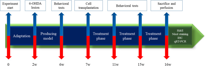
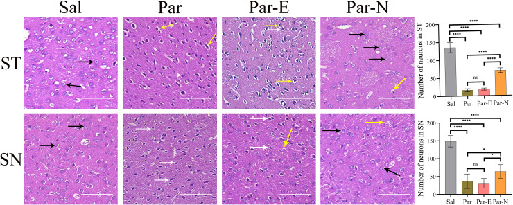
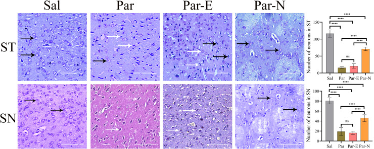
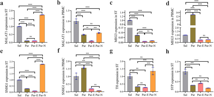
Long non-coding RNAs (lncRNAs) are biomarkers for diagnosis and treatment of Parkinson's disease (PD). Since dopaminergic cell transplantation is a clinical method to treat PD, this study investigated the effects of dopaminergic cell therapy on the expression of some lncRNAs and genes related to PD. In this study, Twenty-eight rats were randomly assigned to four experimental groups. The control group (Sal group) received saline injections. The Par group was a PD rat model with 6-hydroxydopamine (6-OHDA) injection in right striatum (ST). PD animals were transplanted by undifferentiated P19 stem cells (Par-E group), and P19-derived dopaminergic cells (Par-N group). Cell transplant effects were evaluated using behavioral tests (cylinder, open field, and rotarod tests), and histological methods (H&E and Nissl staining, and immunohistochemistry). Moreover, the expression of lncRNAs MALAT1, MEG3, and SNHG1, alongside specific neuronal (synaptophysin) and dopaminergic (tyrosine hydroxylase) markers was evaluated by qRT-PCR. Behavioral and histopathological examinations revealed that cell transplantation partially compensated dopaminergic cell degeneration in ST and substantia nigra (SN) of PD rats. The expression of MALAT1, SNHG1, and MEG3 was decreased in the ST of the Par group, while MEG3 and SNHG1 gene expression was increased in PBMC relative to the Sal group. In PBMC of the Par-N group, all three lncRNAs showed a reduction in their expression. Conversely, MALAT1 and SNHG1 expression was increased in ST tissue, while MEG3 gene expression was decreased compared to the Sal group. In conclusion, dopaminergic cell transplantation could change the lncRNAs expression. Furthermore, it partially improves symptoms in PD rats.


Conflict of interest statement

The authors declare no competing interests.

Figures

Fig. 1
Fig. 1. The experimental design of the present study.
The rats were divided into four main groups. A line of embryonal carcinoma stem cells, P19, was differentiated into dopaminergic neurons and injected into the animals. Behavioral tests were performed both before and after cell transplantation. The experiment lasted 16 weeks. Finally, animals were euthanized and brain samples were collected for qRT-PCR and histopathological analysis.
Fig. 2
Fig. 2. Behavioral tests after unilateral 6-OHDA-induced intrastriatal lesion and cell transplantation.
The following behavior tests including cylinder, rotation, and open field tests were taken from the rats at the time of lesion induction, and 30 and 60 days after the surgery. Statistical analysis exhibited a significant difference between the groups 30 and 60 days after transplantation. a The cylinder test exhibited a significantly increased number of wall touches of the contralateral front paw (the opposite side of the lesion) on the cylinder wall in Par, Par-E, and Par-N groups (n = 7) as compared to the Sal group. On the other hand, the Par-N group relative to Par and Par-E groups exhibited a significant reduction in using the left front paw. b Rotation test diagram after apomorphine administration. Par-N exhibited lower contralateral rotations compared to the Par and Par-E group. c The time spent in the center of the field showed no differences in Par-E and Par groups. These groups had a significant difference compared to the Sal group 60 days after transplantation. d The analysis of the open field-distance traveled test indicated a significant decrease in distance traveled in all the experimental groups compared to the Sal group. e Velocity in the Par, Par-E, and Par-N groups was significantly reduced compared to the Sal group. Nevertheless, the Par-N group displayed a significant increase in contrast to the Par and Par-E group (P < 0.001). f Maps indicative of rat activity after 60 days. All the data of these experiments were mentioned as the mean ± standard deviation (SD) (n = 7). One-way ANOVA was used to evaluate the differences between the mean counts of the groups. P values less than 0.05 (*), 0.01 (**), 0.001 (***), and 0.0001 (****) were regarded as statistically significant. BT before transplantation, AT30 30 days after transplantation, AT60 60 days after transplantation, Sal (control: the rats injected with isotonic saline solution); Par (Parkinson’s rat model: 6-OHDAlesioned animals); Par-E (Parkinson’s rats under cell therapy: A line of embryonal carcinoma stem cells, P19 was transfected with pCMV3-GFPSpark vector and injected into the animals); Par-N (Parkinson’s rats under cell therapy: P19 cells were transfected with pCMV3-NR4A2-GFPSpark vector and injected into the animals).
Fig. 3
Fig. 3. Histological changes in the striatum (ST) and substantia nigra (SN) of the rats, stained with H&E.
The tissue sections of the Sal group exhibited normal structure (black arrows). In Par and Par-E groups, destroyed neurons (white arrows), and neuropil vacuolation (yellow arrows) were observed. The destruction of neurons was reduced in the Par-N group. Some neurons indicated degenerative changes and mild neuropilian vacuolization. Furthermore, the number of neurons in the ST and SN in all groups is shown in the graphs (scale bar = 10 μm). All the data were mentioned as the mean ± standard deviation (SD), (n = 7). One-way ANOVA was used to evaluate the differences between the mean counts of the groups. P values less than 0.05 (*), 0.01 (**), 0.001 (***), and 0.0001 (****) were regarded as statistically significant. Sal control: the rats injected with isotonic saline solution, Par Parkinson’s rat model: 6-OHDA-lesioned animals, Par-E Parkinson’s rats under cell therapy: A line of embryonal carcinoma stem cells, P19 was transfected with pCMV3-GFPSpark vector and injected into the animals, Par-N Parkinson’s rats under cell therapy: P19 cells were transfected with pCMV3-C-NR4A2-GFPSpark vector and injected into the animals.
Fig. 4
Fig. 4. Histological changes in the striatum (ST) and substantia nigra (SN) of rat brain by Nissl staining.
The ST and SN sections of the Sal group exhibited normal structure. In the Par and Par-E groups, the neurons are severely damaged. In the Par-N group, the Nissl substances were increased and the destruction of neurons was significantly reduced. Normal neurons are shown with black arrows and degenerated neurons with white arrows. Effects of 6-OHDA and cell transplantation on the number of neurons in the SN and ST were also shown in graphs (scale bar = 10 μm). All the data were mentioned as the mean ± standard deviation (SD), (n = 7). One-way ANOVA was used to evaluate the differences between the mean counts of the groups. P-values less than 0.05 (*), 0.01 (**), 0.001 (***), and 0.0001 (****) were regarded as statistically significant. Sal control: the rats injected with isotonic saline solution, Par (Parkinson’s rat model: 6-OHDA-lesioned animals); Par-E (Parkinson’s rats under cell therapy: A line of embryonal carcinoma stem cells, P19 was transfected with pCMV3-GFPSpark vector and injected into the animals); Par-N (Parkinson’s rats under cell therapy: P19 cells were transfected with pCMV3-C-NR4A2-GFPSpark vector and injected into the animals).
Fig. 5
Fig. 5. The effect of cell transplantation on the expression of MALAT1, MEG3, SNHG1, TH, and SYP genes in Parkinsonian rats determined by RT-qPCR.
a MALAT1 expression in ST. b MALAT1 expression in PBMC. c MEG3 expression in ST. d MEG3 expression in PBMC. e SNHG1 expression in ST. f SNHG1 expression in PBMC. g TH expression in ST. h SYP expression in ST. All the data were mentioned as the mean ± standard deviation (SD) (n = 7). One-way ANOVA was used to evaluate the differences between the mean counts of the groups. P values less than 0.05 (*), 0.01 (**), 0.001 (***), and 0.0001 (****) were regarded as statistically significant. Sal (control: the rats injected with isotonic saline solution); Par (Parkinson’s rat model: 6-OHDA-lesioned animals); Par-E (Parkinson’s rats under cell therapy: A line of embryonal carcinoma (EC) stem cells, P19 was transfected with pCMV3-GFPSpark vector and injected into the animals); Par-N (Parkinson’s rats under cell therapy: P19 EC cells were transfected with pCMV3-C-NR4A2-GFPSpark vector and injected into the animals.
Fig. 6
Fig. 6. Effect of 6-OHDA injection and cell transplantation on the number of TH+ neurons and cell morphology in the striatum (ST) and substantia nigra (SN) tissues.
The Sal group showed normal neurons in both ST and SN (black arrows). The neurons in the Par group were degenerated due to the injection of 6-OHDA in the ST tissue (white arrows). No significant therapeutic effect was observed in the Par-E group. While, in the Par-N group, the tissues were partially normal (white arrows). The graphs indicate the number of TH-positive neurons in the Sal, Par, Par-E, and Par-N groups (scale bar = 10 μm). All the data were mentioned as the mean ± standard deviation (SD) (n = 7). One-way ANOVA was used to evaluate the differences between the mean counts of the groups. P-values less than 0.05 (*), 0.01 (**), 0.001 (***), and 0.0001 (****) were regarded as statistically significant. Sal control: the rats injected with isotonic saline solution, Par Parkinson’s rat model: 6-OHDA-lesioned animals, Par-E Parkinson’s rats under cell therapy: A line of embryonal carcinoma (EC) stem cells, P19 was transfected with pCMV3-GFPSpark vector and injected into the animals); Par-N (Parkinson’s rats under cell therapy: P19 EC cells were transfected with pCMV3-C-NR4A2-GFPSpark vector and injected into the animals.

Similar articles

Cited by

References

    1. Carvalho MM, et al. Behavioral characterization of the 6-hydroxidopamine model of Parkinson’s disease and pharmacological rescuing of non-motor deficits. Mol. Neurodegener. 2013;8:14. doi: 10.1186/1750-1326-8-14. - DOI - PMC - PubMed
    1. Cai L-J, et al. LncRNA MALAT1 facilitates inflammasome activation via epigenetic suppression of Nrf2 in Parkinson’s disease. Mol. Brain. 2020;13:1–15. doi: 10.1186/s13041-020-00656-8. - DOI - PMC - PubMed
    1. Antoine, B. et al. Whole-brain comparison of rodent and human brains using spatial transcriptomics. Elife11, 10.7554/elife.79418 (2022). - PMC - PubMed
    1. Maegawa, H. & Niwa, H. in Experimental Models of Parkinson’s Disease 95–110 (Springer, 2021).
    1. Xiao Z, et al. The potential therapy with dental tissue-derived mesenchymal stem cells in Parkinson’s disease. Stem Cell Res. Ther. 2021;12:1–11. doi: 10.1186/s13287-020-01957-4. - DOI - PMC - PubMed