. 2024 Mar 12;14(1):5967.
doi: 10.1038/s41598-024-56199-8.

Dual-energy computed tomography and micro-computed tomography for assessing bone regeneration in a rabbit tibia model

Affiliations

Dual-energy computed tomography and micro-computed tomography for assessing bone regeneration in a rabbit tibia model

Danyang Su et al. Sci Rep. .

Abstract

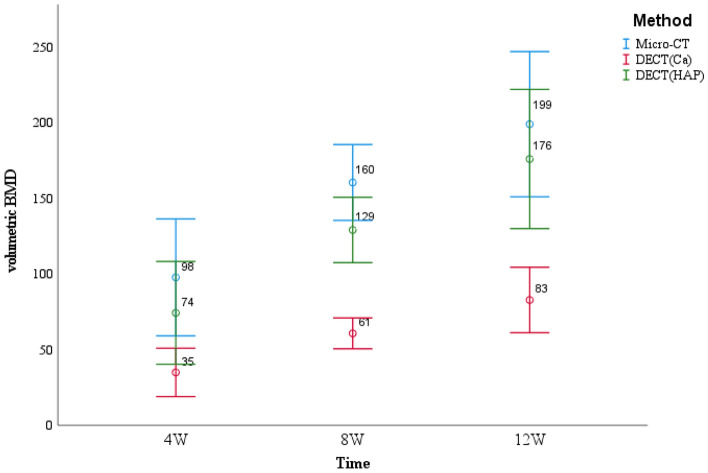
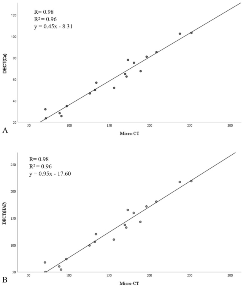
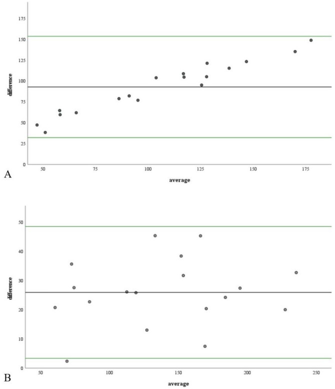
To gain a more meaningful understanding of bone regeneration, it is essential to select an appropriate assessment method. Micro-computed tomography (Micro-CT) is widely used for bone regeneration because it provides a substantially higher spatial resolution. Dual-energy computed tomography (DECT) ensure shorter scan time and lower radiation doses during quantitative evaluation. Therefore, in this study, DECT and Micro-CT were used to evaluate bone regeneration. We created 18 defects in the tibial plateau of the rabbits and filled them with porous polyetheretherketone implants to promote bone regeneration. At 4, 8, and 12 weeks, Micro-CT and DECT were used to assess the bone repair in the defect region. In comparison to Micro-CT (152 ± 54 mg/cm3), the calcium density values and hydroxyapatite density values obtained by DECT [DECT(Ca) and DECT(HAP)] consistently achieved lower values (59 ± 25 mg/cm3, 126 ± 53 mg/cm3). In addition, there was a good association between DECT and Micro-CT (R = 0.98; R2 = 0.96; DECT(Ca): y = 0.45x-8.31; DECT(HAP): y = 0.95x-17.60). This study highlights the need to use two different imaging methods, each with its advantages and disadvantages, to better understand the bone regeneration process.

Keywords: Bone regeneration; Calcium density; DECT; Hydroxyapatite density; Micro-CT; Porous PEEK implants.


Conflict of interest statement

The authors declare no competing interests.

Figures

Figure 1
Figure 1
DECT(Ca), DECT(HAP) and micro-CT were used to obtain the trend of volumetric BMD values in different periods.
Figure 2
Figure 2
Scatter plots with a regression line, correlation coefficient (R), coefficient of determination(R2) and estimated regression model showing the associations between volumetric BMD obtained via Micro-CT and DECT(Ca) (A), as well as volumetric BMD obtained via Micro-CT and DECT(HAP) (B).
Figure 3
Figure 3
Bland–Altman plot of the difference between the measurements obtained by the two methods against their mean value. (A) is for DECT(Ca), and (B) is for DECT(HAP). The black line displays the mean difference, representing the bias between the methods. The green lines indicate the limits of agreement (mean of the differences ± 1.96 SD of the differences).
Figure 4
Figure 4
3D imaging from DECT (AC) and micro-CT (DF) scans after post-processing. In images (AC), the color of the bone defect area gradually changed from gold to white, indicating that the bone repair effect was better. In the three images (DF), the bone appeared gray, and the more grayer deposition, the better the bone repair effect.
Figure 5
Figure 5
Micro-CT (A, C, and E) and DECT (B, D, and F) images of the defect region.

Similar articles

Cited by

References

    1. da Silva Morais A, Oliveira JM, Reis RL. Small animal models. Adv. Exp. Med. Biol. 2018;1059:423–439. doi: 10.1007/978-3-319-76735-2_19. - DOI - PubMed
    1. Bagi CM, Zakur DE, Berryman E, Andresen CJ, Wilkie D. Correlation between μCT imaging, histology and functional capacity of the osteoarthritic knee in the rat model of osteoarthritis. J. Transl. Med. 2015;13:276. doi: 10.1186/s12967-015-0641-7. - DOI - PMC - PubMed
    1. Bauer JS, et al. Multislice-CT for structure analysis of trabecular bone—A comparison with micro-CT and biomechanical strength. Rofo. 2004;176:709–718. doi: 10.1055/s-2004-813078. - DOI - PubMed
    1. Bissinger O, et al. Micro-CT vs. whole body multirow detector CT for analysing bone regeneration in an animal model. PLoS One. 2016;11:e0166540. doi: 10.1371/journal.pone.0166540. - DOI - PMC - PubMed
    1. Bouxsein ML, et al. Guidelines for assessment of bone microstructure in rodents using micro-computed tomography. J. Bone Miner. Res. 2010;25:1468–1486. doi: 10.1002/jbmr.141. - DOI - PubMed