

BMP2 rs1005464 is associated with mandibular condyle size variation
- PMID: 38472272
- PMCID: PMC10933287
- DOI: 10.1038/s41598-024-56530-3
BMP2 rs1005464 is associated with mandibular condyle size variation
Abstract
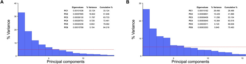
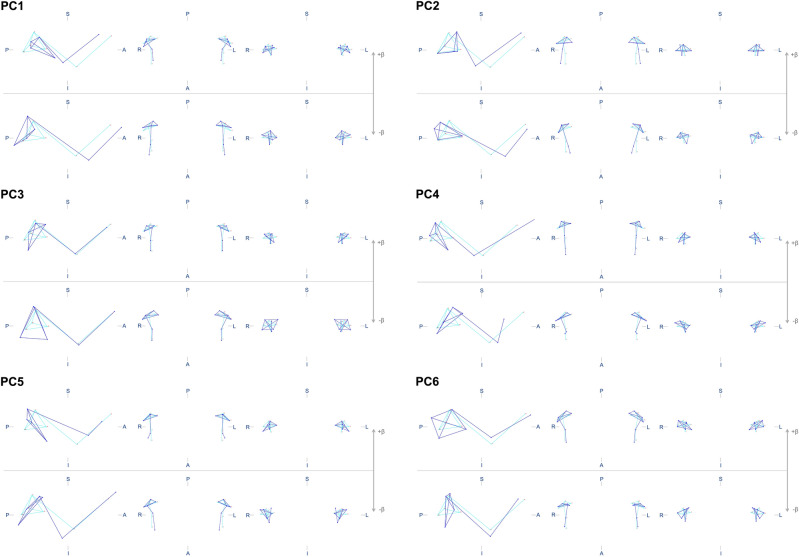
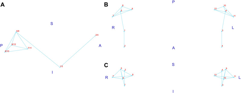
This study aimed to evaluate the association between single nucleotide polymorphisms (SNPs) in endochondral development-related genes and mandibular condyle shape, size, volume, and symmetry traits. Cone-beam Computed Tomographies and genomic DNA from 118 individuals were evaluated (age range: 15-66 years). Data from twelve 3D landmarks on mandibular condyles were submitted to morphometric analyses including Procrustes fit, principal component analysis, and estimation of centroid sizes and fluctuating asymmetry scores. Condylar volumes were additionally measured. Seven SNPs across BMP2, BMP4, RUNX2 and SMAD6 were genotyped. Linear models were fit to evaluate the effect of the SNPs on the mandibular condyles' quantitative traits. Only the association between BMP2 rs1005464 and centroid size remained significant after adjusting to account for the false discovery rate due to multiple testing. Individuals carrying at least one A allele for this SNP showed larger condylar size than common homozygotes GG (β = 0.043; 95% CI: 0.014-0.071; P value = 0.028). The model including BMP2 rs1005464, age and sex of the participants explained 17% of the variation in condylar size. Shape, volume, and symmetry were not associated with the evaluated SNPs. These results suggest that BMP2 rs1005464 might be associated with variation in the mandibular condyles size.
© 2024. The Author(s).
Conflict of interest statement
The authors declare no competing interests.
Figures


Similar articles
-
Investigation of Genetic Polymorphisms in BMP2, BMP4, SMAD6, and RUNX2 and Persistent Apical Periodontitis.J Endod. 2021 Feb;47(2):278-285. doi: 10.1016/j.joen.2020.11.014. Epub 2020 Nov 24. J Endod. 2021. PMID: 33245975
-
Assessing the prevalence of S-shaped root canal and associated genes in humans.Ann Anat. 2022 Oct;244:151977. doi: 10.1016/j.aanat.2022.151977. Epub 2022 Jul 2. Ann Anat. 2022. PMID: 35787440
-
A volumetric study of mandibular condyles in orthognathic patients by semiautomatic segmentation.Oral Maxillofac Surg. 2022 Jun;26(2):205-212. doi: 10.1007/s10006-021-00976-6. Epub 2021 Jun 10. Oral Maxillofac Surg. 2022. PMID: 34114116 Free PMC article.
-
Relationship between occlusal force and mandibular condyle morphology. Evaluated by limited cone-beam computed tomography.Angle Orthod. 2009 Nov;79(6):1063-9. doi: 10.2319/120908-620R.1. Angle Orthod. 2009. PMID: 19852595
-
[Cone-beam CT analysis of vertical control of mandible and changes of temporomandibular joint in adult patients with skeletal class Ⅱ malocclusion with high angle].Zhonghua Kou Qiang Yi Xue Za Zhi. 2022 Nov 9;57(11):1147-1155. doi: 10.3760/cma.j.cn112144-20220301-00086. Zhonghua Kou Qiang Yi Xue Za Zhi. 2022. PMID: 36379894 Chinese.
References
-
- Moss ML, Rankow RM. The role of the functional matrix in mandibular growth. Angle Orthod. 1968;38:95–103. - PubMed
-
- Petrovic AG. Mechanisms and regulation of mandibular condylar growth. Acta Morphol. Neerl. Scand. 1972;10:25–34. - PubMed
-
- Duterloo, H. S. & Wolters, J. M. Experiments on the significance of articular function as a stimulating chondrogenic factor for the growth of secondary cartilages of the rat mandible. Trans. Eur. Orthod. Soc. 103–115 (1971). - PubMed
MeSH terms
Substances
Grants and funding
- 001/Coordenação de Aperfeiçoamento de Pessoal de Nível Superior
- E-26/201337/2022/Fundação Carlos Chagas Filho de Amparo à Pesquisa do Estado do Rio de Janeiro
- E-26/200.199/2023/Fundação Carlos Chagas Filho de Amparo à Pesquisa do Estado do Rio de Janeiro
- Küchler/Kirschneck accepted on July 4th, 2019/Alexander von Humboldt-Stiftung
- Küchler/Kirschneck accepted on July 4th, 2019/Alexander von Humboldt-Stiftung
LinkOut - more resources
Full Text Sources
