

Altered conformational dynamics contribute to species-specific effects of cytochrome c mutations on caspase activation
- PMID: 38472487
- PMCID: PMC11098916
- DOI: 10.1007/s00775-024-02044-2
Altered conformational dynamics contribute to species-specific effects of cytochrome c mutations on caspase activation
Abstract
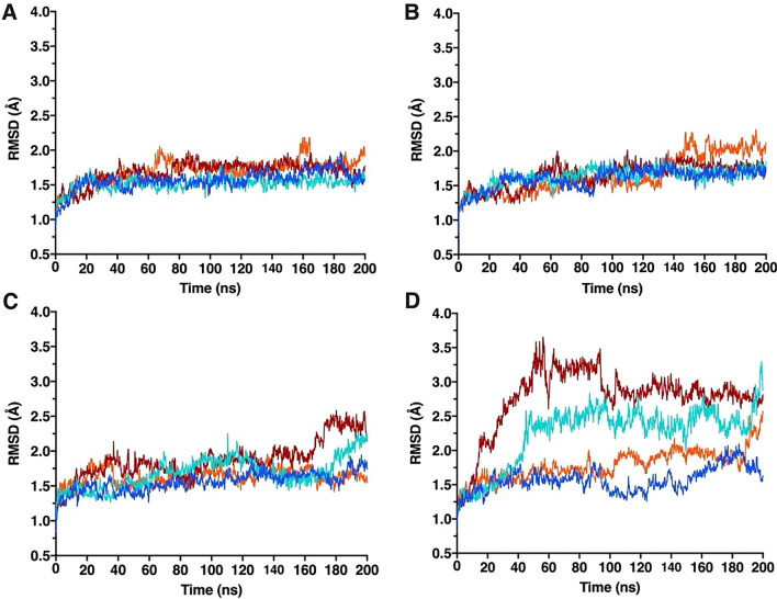
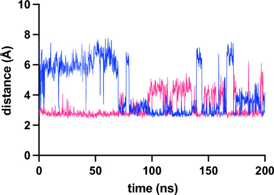
Variants in the gene encoding human cytochrome c (CYCS) cause mild autosomal dominant thrombocytopenia. Despite high sequence conservation between mouse and human cytochrome c, this phenotype is not recapitulated in mice for the sole mutant (G41S) that has been investigated. The effect of the G41S mutation on the in vitro activities of cytochrome c is also not conserved between human and mouse. Peroxidase activity is increased in both mouse and human G41S variants, whereas apoptosome activation is increased for human G41S cytochrome c but decreased for mouse G41S cytochrome c. These apoptotic activities of cytochrome c are regulated at least in part by conformational dynamics of the main chain. Here we use computational and in vitro approaches to understand why the impact of the G41S mutation differs between mouse and human cytochromes c. The G41S mutation increases the inherent entropy and main chain mobility of human but not mouse cytochrome c. Exclusively in human G41S cytochrome c this is accompanied by a decrease in occupancy of H-bonds between protein and heme during simulations. These data demonstrate that binding of cytochrome c to Apaf-1 to trigger apoptosome formation, but not the peroxidase activity of cytochrome c, is enhanced by increased mobility of the native protein conformation.
Keywords: Apaf-1; Apoptosis; Cytochrome c; Molecular dynamics; Peroxidase.
© 2024. The Author(s).
Conflict of interest statement
The authors have no competing interests to declare that are relevant to the content of this article.
Figures


Similar articles
-
Interspecies Variation in the Functional Consequences of Mutation of Cytochrome c.PLoS One. 2015 Jun 18;10(6):e0130292. doi: 10.1371/journal.pone.0130292. eCollection 2015. PLoS One. 2015. PMID: 26086723 Free PMC article.
-
Altered structure and dynamics of pathogenic cytochrome c variants correlate with increased apoptotic activity.Biochem J. 2021 Feb 12;478(3):669-684. doi: 10.1042/BCJ20200793. Biochem J. 2021. PMID: 33480393
-
Conformational change and human cytochrome c function: mutation of residue 41 modulates caspase activation and destabilizes Met-80 coordination.J Biol Inorg Chem. 2013 Mar;18(3):289-97. doi: 10.1007/s00775-012-0973-1. Epub 2013 Jan 19. J Biol Inorg Chem. 2013. PMID: 23334161 Free PMC article.
-
Regulation of apoptosis by the redox state of cytochrome c.Biochim Biophys Acta. 2008 Jul-Aug;1777(7-8):877-81. doi: 10.1016/j.bbabio.2008.03.024. Epub 2008 Apr 3. Biochim Biophys Acta. 2008. PMID: 18439415 Review.
-
Apaf-1/cytochrome c apoptosome: an essential initiator of caspase activation or just a sideshow?Cell Death Differ. 2003 Jan;10(1):16-8. doi: 10.1038/sj.cdd.4401166. Cell Death Differ. 2003. PMID: 12655291 Review. No abstract available.
References
Publication types
MeSH terms
LinkOut - more resources
Full Text Sources
Molecular Biology Databases
Miscellaneous
