

Repeatability of deuterium metabolic imaging of healthy volunteers at 3 T
- PMID: 38472611
- PMCID: PMC10933246
- DOI: 10.1186/s41747-024-00426-4
Repeatability of deuterium metabolic imaging of healthy volunteers at 3 T
Abstract
Background: Magnetic resonance (MR) imaging of deuterated glucose, termed deuterium metabolic imaging (DMI), is emerging as a biomarker of pathway-specific glucose metabolism in tumors. DMI is being studied as a useful marker of treatment response in a scan-rescan scenario. This study aims to evaluate the repeatability of brain DMI.
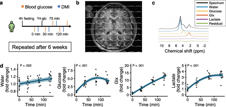
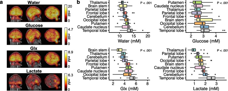
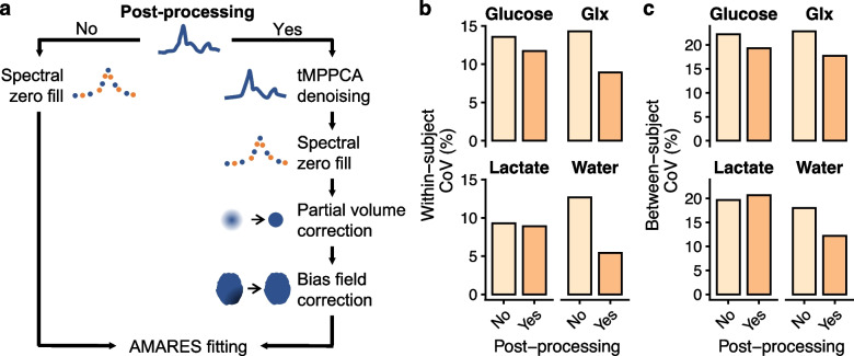
Methods: A repeatability study was performed in healthy volunteers from December 2022 to March 2023. The participants consumed 75 g of [6,6'-2H2]glucose. The delivery of 2H-glucose to the brain and its conversion to 2H-glutamine + glutamate, 2H-lactate, and 2H-water DMI was imaged at baseline and at 30, 70, and 120 min. DMI was performed using MR spectroscopic imaging on a 3-T system equipped with a 1H/2H-tuned head coil. Coefficients of variation (CoV) were computed for estimation of repeatability and between-subject variability. In a set of exploratory analyses, the variability effects of region, processing, and normalization were estimated.
Results: Six male participants were recruited, aged 34 ± 6.5 years (mean ± standard deviation). There was 42 ± 2.7 days between sessions. Whole-brain levels of glutamine + glutamate, lactate, and glucose increased to 3.22 ± 0.4 mM, 1.55 ± 0.3 mM, and 3 ± 0.7 mM, respectively. The best signal-to-noise ratio and repeatability was obtained at the 120-min timepoint. Here, the within-subject whole-brain CoVs were -10% for all metabolites, while the between-subject CoVs were -20%.
Conclusions: DMI of glucose and its downstream metabolites is feasible and repeatable on a clinical 3 T system.
Trial registration: ClinicalTrials.gov, NCT05402566 , registered the 25th of May 2022.
Relevance statement: Brain deuterium metabolic imaging of healthy volunteers is repeatable and feasible at clinical field strengths, enabling the study of shifts in tumor metabolism associated with treatment response.
Key points: • Deuterium metabolic imaging is an emerging tumor biomarker with unknown repeatability. • The repeatability of deuterium metabolic imaging is on par with FDG-PET. • The study of deuterium metabolic imaging in clinical populations is feasible.
Keywords: Biomarkers (tumor); Brain; Deuterium; Glucose; Magnetic resonance imaging.
© 2024. The Author(s).
Conflict of interest statement
MV and RFS are employees of GE Healthcare. The non-industry authors were in complete control of the data and information presented in the study.
Figures

Similar articles
-
Deuterium Metabolic Imaging of Alzheimer Disease at 3-T Magnetic Field Strength: A Pilot Case-Control Study.Radiology. 2024 Jul;312(1):e232407. doi: 10.1148/radiol.232407. Radiology. 2024. PMID: 39012255 Free PMC article.
-
Noninvasive 3-Dimensional 1 H-Magnetic Resonance Spectroscopic Imaging of Human Brain Glucose and Neurotransmitter Metabolism Using Deuterium Labeling at 3T : Feasibility and Interscanner Reproducibility.Invest Radiol. 2023 Jun 1;58(6):431-437. doi: 10.1097/RLI.0000000000000953. Epub 2023 Feb 4. Invest Radiol. 2023. PMID: 36735486 Free PMC article.
-
Balanced Steady-State Free Precession Enables High-Resolution Dynamic 3D Deuterium Metabolic Imaging of the Human Brain at 7T.Invest Radiol. 2025 Apr 25:10.1097/RLI.0000000000001196. doi: 10.1097/RLI.0000000000001196. Online ahead of print. Invest Radiol. 2025. PMID: 40273422
-
Advances and prospects in deuterium metabolic imaging (DMI): a systematic review of in vivo studies.Eur Radiol Exp. 2024 Jun 3;8(1):65. doi: 10.1186/s41747-024-00464-y. Eur Radiol Exp. 2024. PMID: 38825658 Free PMC article.
-
Falls prevention interventions for community-dwelling older adults: systematic review and meta-analysis of benefits, harms, and patient values and preferences.Syst Rev. 2024 Nov 26;13(1):289. doi: 10.1186/s13643-024-02681-3. Syst Rev. 2024. PMID: 39593159 Free PMC article.
Cited by
-
PyAMARES, an Open-Source Python Library for Fitting Magnetic Resonance Spectroscopy Data.Diagnostics (Basel). 2024 Nov 27;14(23):2668. doi: 10.3390/diagnostics14232668. Diagnostics (Basel). 2024. PMID: 39682576 Free PMC article.
-
Open-source implementation of X-nuclear sequences using the Pulseq framework.Magn Reson Med. 2025 Aug;94(2):651-664. doi: 10.1002/mrm.30509. Epub 2025 Apr 2. Magn Reson Med. 2025. PMID: 40173321
-
Test-Retest Reproducibility of Reduced-Field-of-View Density-Weighted CRT MRSI at 3T.Tomography. 2024 Mar 29;10(4):493-503. doi: 10.3390/tomography10040038. Tomography. 2024. PMID: 38668396 Free PMC article.
References
Publication types
MeSH terms
Substances
Associated data
LinkOut - more resources
Full Text Sources
Medical