

Exosomes Derived from Mouse Breast Carcinoma Cells Facilitate Diabetic Wound Healing
- PMID: 38472732
- PMCID: PMC11087414
- DOI: 10.1007/s13770-024-00629-1
Exosomes Derived from Mouse Breast Carcinoma Cells Facilitate Diabetic Wound Healing
Abstract
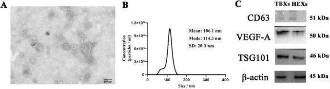
Background: Exosomes derived from breast cancer have been reported to play a role in promoting cell proliferation, migration, and angiogenesis, which has the potential to accelerate the healing process of diabetic wounds. The aim of this investigation was to examine the function of exosomes originating from 4T1 mouse breast carcinoma cells (TEXs) in the process of diabetic wound healing.
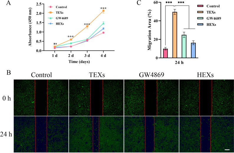
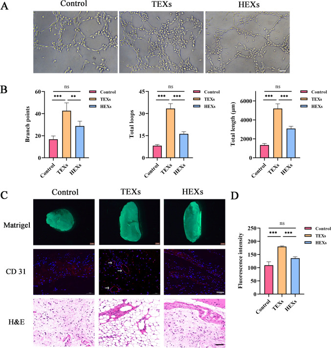
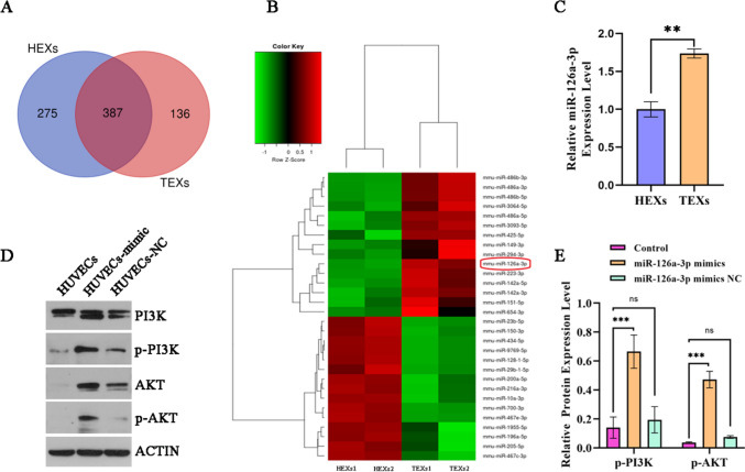
Methods: The assessment of primary mouse skin fibroblasts cell proliferation and migration was conducted through the utilization of CCK-8 and wound healing assays, while the tube formation of HUVECs was evaluated by tube formation assay. High-throughput sequencing, RT-qPCR and cell experiments were used to detect the roles of miR-126a-3p in HUVECs functions in vitro. The in vivo study employed a model of full-thickness excisional wounds in diabetic subjects to explore the potential therapeutic benefits of TEXs. Immunohistochemical and immunofluorescent techniques were utilized to evaluate histological changes in skin tissues.
Results: The findings suggested that TEXs facilitate diabetic wound healing through the activation of cell migration, proliferation, and angiogenesis. An upregulation of miR-126a-3p has been observed in TEXs, and it has demonstrated efficient transferability from 4T1 cells to HUVEC cells. The activation of the PI3K/Akt pathway has been attributed to miR-126a-3p derived from TEXs.
Conclusions: The promotion of chronic wound healing can be facilitated by TEXs through the activation of cellular migration, proliferation, and angiogenesis. The activation of the PI3K/Akt pathway by miR-126a-3p originating from TEXs has been discovered, indicating a potential avenue for enhancing the regenerative capabilities of wounds treated with TEXs.
Keywords: Angiogenesis; Cell proliferation and migration; Exosomes; Wound healing; miR-126a-3p.
© 2024. Korean Tissue Engineering and Regenerative Medicine Society.
Conflict of interest statement
The authors report no conflicts of interest in this work.
Figures
Similar articles
-
Empagliflozin-Pretreated MSC-Derived Exosomes Enhance Angiogenesis and Wound Healing via PTEN/AKT/VEGF Pathway.Int J Nanomedicine. 2025 Apr 22;20:5119-5136. doi: 10.2147/IJN.S512074. eCollection 2025. Int J Nanomedicine. 2025. PMID: 40297404 Free PMC article.
-
Exosomes derived from atorvastatin-pretreated MSC accelerate diabetic wound repair by enhancing angiogenesis via AKT/eNOS pathway.Stem Cell Res Ther. 2020 Aug 12;11(1):350. doi: 10.1186/s13287-020-01824-2. Stem Cell Res Ther. 2020. PMID: 32787917 Free PMC article.
-
IFN-γ enhances the therapeutic efficacy of MSCs-derived exosome via miR-126-3p in diabetic wound healing by targeting SPRED1.J Diabetes. 2024 Jan;16(1):e13465. doi: 10.1111/1753-0407.13465. Epub 2023 Aug 30. J Diabetes. 2024. PMID: 37646268 Free PMC article.
-
[Research advances on the role of microRNA engineered exosomes in diabetic wounds].Zhonghua Shao Shang Yu Chuang Mian Xiu Fu Za Zhi. 2024 Feb 20;40(2):190-195. doi: 10.3760/cma.j.cn501225-20230721-00011. Zhonghua Shao Shang Yu Chuang Mian Xiu Fu Za Zhi. 2024. PMID: 38418181 Free PMC article. Review. Chinese.
-
Update on Exosomes in Aesthetics.Dermatol Surg. 2022 Aug 1;48(8):862-865. doi: 10.1097/DSS.0000000000003487. Epub 2022 May 17. Dermatol Surg. 2022. PMID: 35580250 Review.
References
Publication types
MeSH terms
Substances
Grants and funding
- 31600791/National Natural Science Foundation of China
- 222102310185/Scientific and Technological Research Project of Science and Technology Department of Henan Province
- 232102311096/Scientific and Technological Research Project of Science and Technology Department of Henan Province
- KFKTYB202117/Open Project Program of the Third Affiliated Hospital of Xinxiang Medical University
- KFKTZD202102/Open Project Program of the Third Affiliated Hospital of Xinxiang Medical University
LinkOut - more resources
Full Text Sources