

Association of Blood NK Cell Phenotype with the Severity of Liver Fibrosis in Patients with Chronic Viral Hepatitis C with Genotype 1 or 3
- PMID: 38472945
- PMCID: PMC10930504
- DOI: 10.3390/diagnostics14050472
Association of Blood NK Cell Phenotype with the Severity of Liver Fibrosis in Patients with Chronic Viral Hepatitis C with Genotype 1 or 3
Abstract
Background: NK cells phenotype and functional state in different genotypes of chronic viral hepatitis C (CVHC), depending on liver fibrosis severity, have not been sufficiently studied, which limits the possibilities for the development of pathology therapy.
Methods: The CVHC diagnosis was based on the EASL recommendations (2018). Clinical examination with liver elastometry was performed in 297 patients with genotype 1 and in 231 patients with genotype 3 CVHC. The blood NK cells phenotype was determined by flow cytometry in 74 individuals with genotype 1 and in 69 individuals with genotype 3 CVHC.
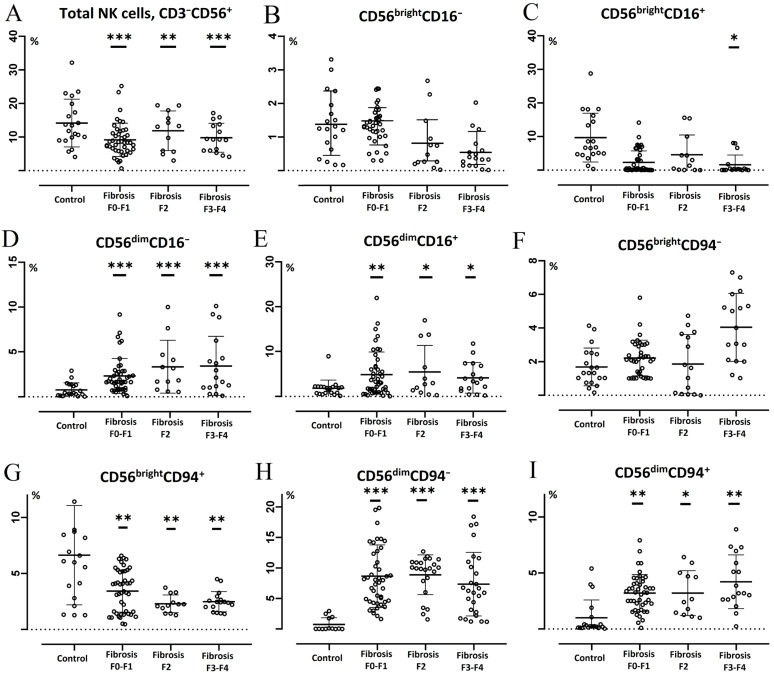
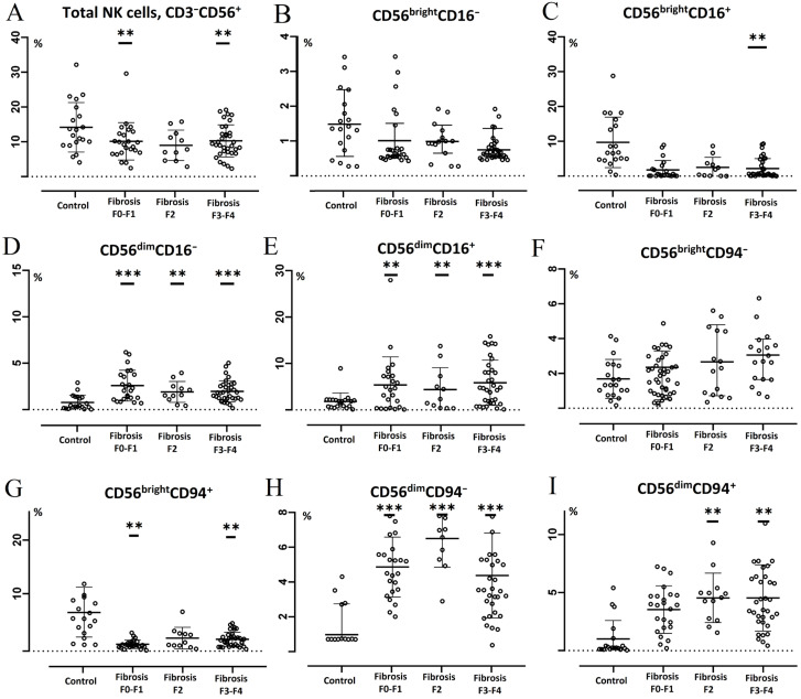
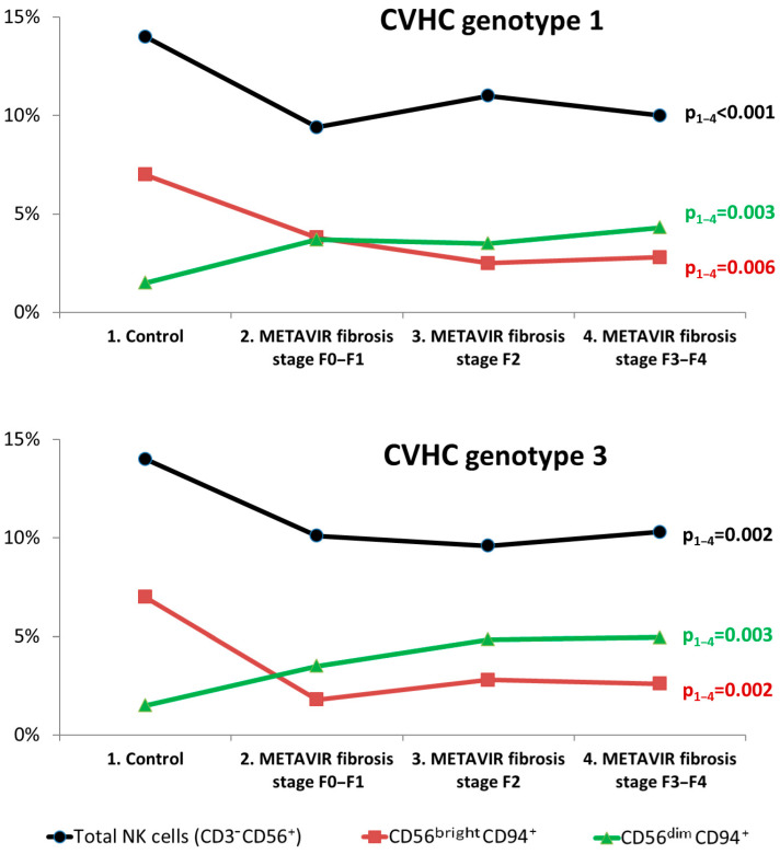
Results: The frequency of METAVIR liver fibrosis stages F3-F4 was 32.5% in individuals with genotype 3, and 20.5% in individuals with genotype 1 CVHC (p = 0.003). In patients with both genotype 1 and genotype 3 CVHC, a decrease in the total number of blood NK cells, CD56brightCD16+ NK cells and an increase in the proportion of CD56dimCD16+ NK cells, CD94+ and CD38 + CD73+ NK cells were registered in patients with fibrosis stage F3-F4 by METAVIR in comparison with persons with METAVIR fibrosis stage F0-F1.
Conclusions: In patients with both genotype 1 and genotype 3 CVHC, an imbalance in the ratio between cytokine-producing and cytotoxic NK cells and an increase in the content of NK cells that express inhibitory molecules were determined in patients with severe liver fibrosis.
Keywords: NK cells; liver fibrosis; phenotype; receptor expression; subsets; viral hepatitis C.
Conflict of interest statement
The authors declare no conflicts of interest.
Figures
Similar articles
-
Liver cirrhosis in HIV/HCV-coinfected individuals is related to NK cell dysfunction and exhaustion, but not to an impaired NK cell modulation by CD4+ T-cells.J Int AIDS Soc. 2019 Sep;22(9):e25375. doi: 10.1002/jia2.25375. J Int AIDS Soc. 2019. PMID: 31536177 Free PMC article.
-
Association of NK Cells with the Severity of Fibrosis in Patients with Chronic Hepatitis C.Diagnostics (Basel). 2023 Jun 27;13(13):2187. doi: 10.3390/diagnostics13132187. Diagnostics (Basel). 2023. PMID: 37443584 Free PMC article.
-
Selective depletion of CD56(dim) NK cell subsets and maintenance of CD56(bright) NK cells in treatment-naive HIV-1-seropositive individuals.J Clin Immunol. 2002 May;22(3):176-83. doi: 10.1023/a:1015476114409. J Clin Immunol. 2002. PMID: 12078859
-
Human Circulating and Tissue-Resident CD56(bright) Natural Killer Cell Populations.Front Immunol. 2016 Jun 30;7:262. doi: 10.3389/fimmu.2016.00262. eCollection 2016. Front Immunol. 2016. PMID: 27446091 Free PMC article. Review.
-
Role of chemokines in the biology of natural killer cells.Curr Top Microbiol Immunol. 2010;341:37-58. doi: 10.1007/82_2010_20. Curr Top Microbiol Immunol. 2010. PMID: 20369317 Review.
References
-
- Bartenschlager R., Baumert T.F., Bukh J., Houghton M., Lemon S.M., Lindenbach B.D., Lohmann V., Moradpour D., Pietschmann T., Rice C.M., et al. Critical challenges and emerging opportunities in hepatitis C virus research in an era of potent antiviral therapy: Considerations for scientists and funding agencies. Virus Res. 2018;248:53–62. doi: 10.1016/j.virusres.2018.02.016. - DOI - PubMed
LinkOut - more resources
Full Text Sources
Research Materials
Miscellaneous
