

Evaluation of Pulmonary Nodules by Radiologists vs. Radiomics in Stand-Alone and Complementary CT and MRI
- PMID: 38472955
- PMCID: PMC10930396
- DOI: 10.3390/diagnostics14050483
Evaluation of Pulmonary Nodules by Radiologists vs. Radiomics in Stand-Alone and Complementary CT and MRI
Abstract
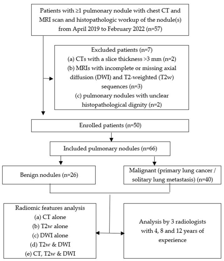
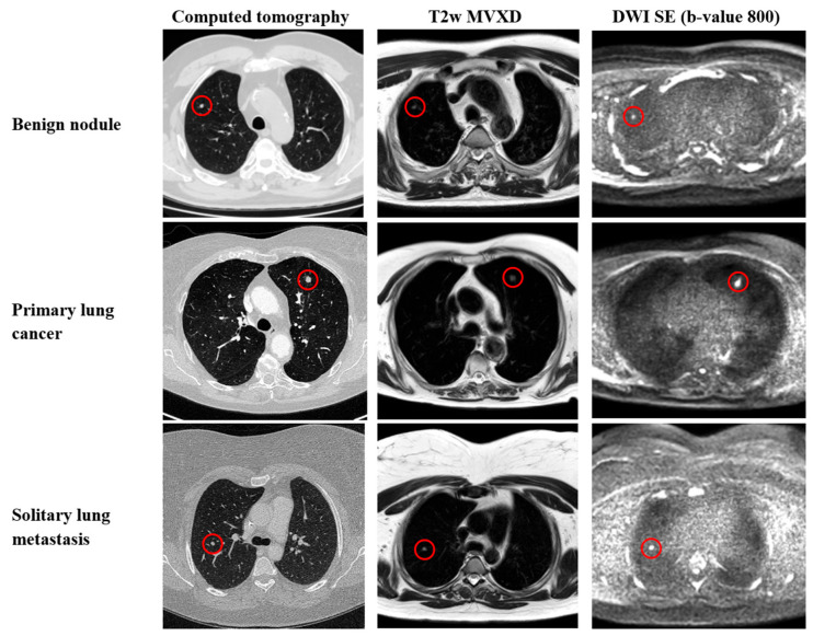
Increased attention has been given to MRI in radiation-free screening for malignant nodules in recent years. Our objective was to compare the performance of human readers and radiomic feature analysis based on stand-alone and complementary CT and MRI imaging in classifying pulmonary nodules. This single-center study comprises patients with CT findings of pulmonary nodules who underwent additional lung MRI and whose nodules were classified as benign/malignant by resection. For radiomic features analysis, 2D segmentation was performed for each lung nodule on axial CT, T2-weighted (T2w), and diffusion (DWI) images. The 105 extracted features were reduced by iterative backward selection. The performance of radiomics and human readers was compared by calculating accuracy with Clopper-Pearson confidence intervals. Fifty patients (mean age 63 +/- 10 years) with 66 pulmonary nodules (40 malignant) were evaluated. ACC values for radiomic features analysis vs. radiologists based on CT alone (0.68; 95%CI: 0.56, 0.79 vs. 0.59; 95%CI: 0.46, 0.71), T2w alone (0.65; 95%CI: 0.52, 0.77 vs. 0.68; 95%CI: 0.54, 0.78), DWI alone (0.61; 95%CI:0.48, 0.72 vs. 0.73; 95%CI: 0.60, 0.83), combined T2w/DWI (0.73; 95%CI: 0.60, 0.83 vs. 0.70; 95%CI: 0.57, 0.80), and combined CT/T2w/DWI (0.83; 95%CI: 0.72, 0.91 vs. 0.64; 95%CI: 0.51, 0.75) were calculated. This study is the first to show that by combining quantitative image information from CT, T2w, and DWI datasets, pulmonary nodule assessment through radiomics analysis is superior to using one modality alone, even exceeding human readers' performance.
Keywords: CT; MRI; artificial intelligence; pulmonary nodule; radiomics.
Conflict of interest statement
The authors declare no conflicts of interest.
Figures
Similar articles
-
Magnetic resonance radiomic feature performance in pulmonary nodule classification and impact of segmentation variability on radiomics.Br J Radiol. 2022 Dec 1;95(1140):20220230. doi: 10.1259/bjr.20220230. Epub 2022 Nov 15. Br J Radiol. 2022. PMID: 36367095 Free PMC article.
-
[Value of radiomics models based on MRI diffusion weighted imaging and apparent diffusion coefficient in differentiating benign and malignant thyroid nodules].Zhonghua Yi Xue Za Zhi. 2023 Nov 7;103(41):3279-3286. doi: 10.3760/cma.j.cn112137-20230913-00453. Zhonghua Yi Xue Za Zhi. 2023. PMID: 37926572 Chinese.
-
Differentiating inflammatory and malignant pulmonary lesions on 3T lung MRI with radiomics of apparent diffusion coefficient maps and T2w derived radiomic feature maps.J Thorac Dis. 2024 May 31;16(5):2875-2893. doi: 10.21037/jtd-23-1456. Epub 2024 May 24. J Thorac Dis. 2024. PMID: 38883623 Free PMC article.
-
Radiomics of pulmonary nodules and lung cancer.Transl Lung Cancer Res. 2017 Feb;6(1):86-91. doi: 10.21037/tlcr.2017.01.04. Transl Lung Cancer Res. 2017. PMID: 28331828 Free PMC article. Review.
-
Deep neural network pulmonary nodule segmentation methods for CT images: Literature review and experimental comparisons.Comput Biol Med. 2023 Sep;164:107321. doi: 10.1016/j.compbiomed.2023.107321. Epub 2023 Aug 9. Comput Biol Med. 2023. PMID: 37595518 Review.
Cited by
-
Auxiliary Diagnosis of Pulmonary Nodules' Benignancy and Malignancy Based on Machine Learning: A Retrospective Study.J Multidiscip Healthc. 2025 Jun 27;18:3735-3748. doi: 10.2147/JMDH.S518166. eCollection 2025. J Multidiscip Healthc. 2025. PMID: 40600198 Free PMC article.
References
-
- MacMahon H., Naidich D.P., Goo J.M., Lee K.S., Leung A.N.C., Mayo J.R., Mehta A.C., Ohno Y., Powell C.A., Prokop M., et al. Guidelines for Management of Incidental Pulmonary Nodules Detected on CT Images: From the Fleischner Society 2017. Radiology. 2017;284:228–243. doi: 10.1148/radiol.2017161659. - DOI - PubMed
LinkOut - more resources
Full Text Sources
