The Vertebrobasilar Trunk and Its Anatomical Variants: A Microsurgical Anatomical Study
- PMID: 38473006
- PMCID: PMC10930859
- DOI: 10.3390/diagnostics14050534
The Vertebrobasilar Trunk and Its Anatomical Variants: A Microsurgical Anatomical Study
Abstract
Background: The trunk of the basilar artery has not been included in microanatomy studies. Anatomical variants of the perforant branches of the vertebrobasilar trunk and their relationship with neural structures are very important in surgical approaches. Surgical dissection for the treatment of vascular lesions requires a perfect knowledge of the microsurgical anatomy.
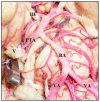
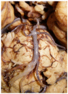
Methods: We conducted a descriptive analysis of 50 brains, which were fixed with formalin at 10% for 2 weeks, and the arterial system was injected with colored latex. After microsurgical dissection, it was divided into three segments: the lower portion went from the anterior spinal artery to the anteroinferior cerebellar artery, the middle segment was raised from the upper limit of the lower portion to the origin of the superior cerebellar artery, and the upper segment ranged from the previous portion until the origin of the posterior cerebral artery.
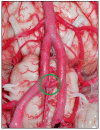
Results: The basilar artery had an average length of 30 mm. The average diameter at its junction with the vertebral arteries was 4.05 mm. The average middle segment was 3.4 mm in diameter and 15.2 mm in length. The diameter of the upper segment was 4.2 mm, and its average length was 3.6 mm. The average number of bulbar arteries was three, and their average diameter was 0. 66 mm. The number of caudal perforator arteries were five on average, with a diameter of 0.32 mm. We found three rare cases of anatomical variants in the vertebra-basilar junction.
Conclusions: The basilar artery emits penetrating branches in its lower, middle, and upper portions. The origin of penetrating branches was single or divided after forming a trunk. However, we observed long branches from perforant arteries.
Keywords: anatomy; arterial trunk; basilar artery; laboratory; neurosurgery; perforants; terminal branches; vertebral artery.
Conflict of interest statement
The authors declare no conflicts of interest.
Figures


Similar articles
-
Novel classification and microsurgical anatomy of the basilar artery: a cadaveric study.Surg Radiol Anat. 2025 Apr 2;47(1):111. doi: 10.1007/s00276-025-03612-0. Surg Radiol Anat. 2025. PMID: 40175830
-
The segmentation of the posterior cerebral artery: a microsurgical anatomic study.Neurosurg Rev. 2019 Mar;42(1):155-161. doi: 10.1007/s10143-018-0972-y. Epub 2018 Apr 6. Neurosurg Rev. 2019. PMID: 29623480
-
Microsurgical anatomy of the intracranial part of the vertebral artery.Neurol Res. 1994 Jun;16(3):171-80. doi: 10.1080/01616412.1994.11740221. Neurol Res. 1994. PMID: 7936084
-
Microsurgical Neurovascular Anatomy of the Brain: The Posterior Circulation (Part II).Acta Biomed. 2021 Aug 26;92(S4):e2021413. doi: 10.23750/abm.v92iS4.12119. Acta Biomed. 2021. PMID: 34437362 Free PMC article. Review.
-
Microsurgical anatomy of the posterior choroidal arteries.Neurol Res. 1995 Oct;17(5):334-44. Neurol Res. 1995. PMID: 8584123 Review.
Cited by
-
Morphometric and Clinical Analysis of the Azygos Anterior Cerebral Artery: Insights From a Cadaveric Case Report Study.Cureus. 2025 Apr 27;17(4):e83063. doi: 10.7759/cureus.83063. eCollection 2025 Apr. Cureus. 2025. PMID: 40432638 Free PMC article.
-
Association of the Middle Accessory Cerebral Artery With Bihemispheric Anterior Cerebral Artery and Median Artery of the Corpus Callosum.Cureus. 2025 Jun 25;17(6):e86733. doi: 10.7759/cureus.86733. eCollection 2025 Jun. Cureus. 2025. PMID: 40718333 Free PMC article.
-
The clinical significance of artery of Percheron in cerebral ischemia: a summary overview.Ann Med Surg (Lond). 2025 May 30;87(7):4310-4315. doi: 10.1097/MS9.0000000000003439. eCollection 2025 Jul. Ann Med Surg (Lond). 2025. PMID: 40851980 Free PMC article. Review.
-
Microsurgical insights: A comprehensive anatomical study of Heubner's recurrent artery.Surg Neurol Int. 2025 May 2;16:158. doi: 10.25259/SNI_81_2025. eCollection 2025. Surg Neurol Int. 2025. PMID: 40469350 Free PMC article.
-
The trinity of the internal carotid artery: Unifying terminologies of the main classifications to improve its surgical understanding.Surg Neurol Int. 2025 May 16;16:177. doi: 10.25259/SNI_27_2025. eCollection 2025. Surg Neurol Int. 2025. PMID: 40469375 Free PMC article.
References
LinkOut - more resources
Full Text Sources

