

Bilateral Renal Ectopia-Prenatal Diagnosis
- PMID: 38473011
- PMCID: PMC10931438
- DOI: 10.3390/diagnostics14050539
Bilateral Renal Ectopia-Prenatal Diagnosis
Abstract
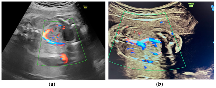
This report explores the diverse spectrum of congenital anomalies of the kidney and urinary tract (CAKUT), ranging from asymptomatic presentations to the most severe form characterized by bilateral renal agenesis. Genitourinary anomalies, a prevalent subset within this domain, account for a significant proportion, constituting 15-20% of anomalies identified during prenatal screening. An ectopic kidney is defined by the presence of an empty renal fossa and the displacement of the kidney from the lumbar region to alternative locations, with the pelvic region emerging as the most prevalent site. The reported case involves bilateral renal ectopia with unilateral duplex kidney. Initial suspicions of a renal anomaly arose during the first trimester, leading to a definitive diagnosis in the second trimester. The patient underwent regular monitoring every four weeks, ultimately delivering a healthy baby at term. This case underscores the frequency of renal anomalies, emphasizing that a considerable proportion remains asymptomatic. These findings contribute to a broader understanding of congenital renal anomalies, their varied manifestations, and the importance of vigilant prenatal screening for early detection and management.
Keywords: duplex kidney; renal anomalies; renal ectopia.
Conflict of interest statement
The authors declare no conflicts of interest.
Figures


References
-
- Duplex Collecting System (by Jones, J.; Niknejad, M.; Guan, H. et al.) [(accessed on 28 January 2024)]. Available online: https://radiopaedia.org.
LinkOut - more resources
Full Text Sources
