. 2024 Feb 22;16(5):877.
doi: 10.3390/cancers16050877.

Preclinical Characterization of the Anti-Leukemia Activity of the CD33/CD16a/NKG2D Immune-Modulating TriNKET® CC-96191

Affiliations

Preclinical Characterization of the Anti-Leukemia Activity of the CD33/CD16a/NKG2D Immune-Modulating TriNKET® CC-96191

Margaret C Lunn-Halbert et al. Cancers (Basel). .

Abstract

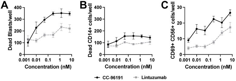
Increasing efforts are focusing on natural killer (NK) cell immunotherapies for AML. Here, we characterized CC-96191, a novel CD33/CD16a/NKG2D immune-modulating TriNKET®. CC-96191 simultaneously binds CD33, NKG2D, and CD16a, with NKG2D and CD16a co-engagement increasing the avidity for, and activation of, NK cells. CC-96191 was broadly active against human leukemia cells in a strictly CD33-dependent manner, with maximal efficacy requiring the co-engagement of CD16a and NKG2D. A frequent CD33 single nucleotide polymorphism, R69G, reduced CC-96191 potency but not maximal activity, likely because of reduced CD33 binding. Similarly, the potency, but not the maximal activity, of CC-96191 was reduced by high concentrations of soluble CD33; in contrast, the soluble form of the NKG2D ligand MICA did not impact activity. In the presence of CD33+ AML cells, CC-96191 activated NK cells but not T cells; while maximum anti-AML efficacy was similar, soluble cytokine levels were 10- to >100-fold lower than with a CD33/CD3 bispecific antibody. While CC-96191-mediated cytolysis was not affected by ABC transporter proteins, it was reduced by anti-apoptotic BCL-2 family proteins. Finally, in patient marrow specimens, CC-96191 eliminated AML cells but not normal monocytes, suggesting selectivity of TriNKET-induced cytotoxicity toward neoplastic cells. Together, these findings support the clinical exploration of CC-96191 as in NCT04789655.

Keywords: CD33; acute myeloid leukemia (AML); immunotherapy; natural killer (NK) cell; trispecific antibody.


Conflict of interest statement

M.T.O., H.C., H.J.T., H.K.J, M.A.J., and J.L. are employees of Bristol Myers Squibb. A.F.C, G.P.C, T.D., D.F., and A.G. are employees of Dragonfly Therapeutics. E.R.A. received honoraria for educational activities and/or conferences and/or travel grants from AbbVie, Astellas, Gilead, and Jazz. R.B.W. received laboratory research grants and/or clinical trial support from Aptevo, Celgene/Bristol Myers Squibb, ImmunoGen, Janssen, Jazz, Kite, Kura, Pfizer, and VOR; has ownership interests in Amphivena; and is (or has been) a consultant to Abbvie, Adicet, Amphivena, BerGenBio, Celgene/Bristol Myers Squibb, GlaxoSmithKline, ImmunoGen, Kura, Orum, and Wugen. The other authors declare no competing financial interests.

Figures

Figure 1
Figure 1
Binding of CC-96191 to CD33, NKG2D, and CD16a. (A) SPR analysis of the binding of CC-96191 or trastuzumab (analyte) to human CD16a (158F), human NKG2D, or combined CD16a and NKG2D. NKG2D alone (12.3 μg/mL), CD16a alone (9 μg/mL), or the mixture of NKG2D and CD16a at the same concentrations each were amine-coupled to the surface of a CM5 Series S Biacore chip. A total of 1.8 μM CC-96191 or trastuzumab were injected for 150 s at 10 μL/min. Dissociation phase was observed for 180 s when regeneration was not needed and 1800 s when natural regeneration of the surface (almost complete dissociation of analyte) was needed between the cycles at the same flow rate. (B) SPR analysis of the sequential binding of saturating concentrations of CD33 (100 nM) and NKG2D (7000 nM) in both orientations across the surface of a Biacore CM5 chip onto which CC-96191 was Fc-captured, with calculation of relative binding stoichiometries. Note: A mixed solution of CD33 and NKG2D was included in the second injection of the first sensorgram to maintain a saturating concentration of NKG2D due to the fast dissociation of NKG2D from CC-96191. (C) SPR analysis of the sequential binding of mFc-NKG2D (7000 nM) and CD16a (3000 nM) when injected in sequential order across CC-96191 when captured via CD33-His that was amine coupled to a Biacore CM5 chip.
Figure 2
Figure 2
Impact of individual binding arms on CC-96191-induced cytotoxicity. (A,C) NK cells from healthy donors were incubated for 2 to 3 h with BATDA-labeled CD33+ EOL-1 target cells at an effector:target (E:T) cell ratio of 5:1 in the presence or absence of (A) CC-96191, CC-96191FC-SILENT, CD33 mAb I07, or CC-96191FC-SILENT and CD33 mAb I07 or (C) CC-96191, CC-96191ΔNKG2D, or CC-96191ΔCD33. (B) CD33+ MOLM-13 or TF-1 cells were incubated with parental (CD16a-) KYHG-1 or KHYG-1CD16a cells at an E:T cell ratio of 5:1 with various concentrations of CC-96191. After 2 days, cell numbers and the percentage of dead cells were quantified by flow cytometry. Data are presented as mean ± SEM from 3 independent experiments performed in duplicate wells. (D) CD33+ target cells (EOL-1, ML-1, TF-1) were incubated with KHYG-1CD16a cells at an E:T cell ratio of 3:1 in the presence or absence of CC-96191, CC-96191ΔNKG2D, or CC-96191ΔCD33 at 1 or 5 nM as indicated. After 2 days, cell numbers and the percentage of dead cells were quantified by flow cytometry. Data are presented as mean ± SEM from 3 independent experiments performed in duplicate wells.
Figure 3
Figure 3
Relationship between CD33 expression and CC-96191-induced cytotoxicity. Sublines of (A) KG-1, (B) ML-1, (C) OCI-AML3, and (D) TF-1 cells, genetically engineered to express different levels of CD33 (as determined by QuantiBRITE), were incubated with KHYG-1CD16a cells at an E:T cell ratio of 3:1 in the presence or absence of CC-96191 at various concentrations as indicated. After 2 days, cell numbers and the percentage of dead cells were quantified by flow cytometry. Data are presented as mean ± SEM from 3 independent experiments performed in duplicate wells.
Figure 4
Figure 4
Impact of CD33 SNPs on CC-96191-induced cytotoxicity. (A,B) MOLM-13 and (C,D) EOL-1 cells with CRISPR/Cas9-induced deletion of CD33 (CD33KO) and sublines in which either full-length wild-type CD33 (WT) or full-length CD33 A14V, R69G, or G304R were overexpressed via lentivirus to a similar degree were incubated with KHYG-1CD16a cells at an E:T cell ratio of 3:1 in the presence or absence of CC-96191 (1–500 pM) as indicated. (E,F) REH cells were transfused with either wild-type CD33 (WT) or full-length CD33 R69G and incubated with KHYG-1CD16a cells at an E:T cell ratio of 3:1 in the presence or absence of CC-96191 (1–500 pM) as indicated. After 2 days, cell numbers and the percentage of dead cells were quantified by flow cytometry. Data are presented as mean ± SEM from 3–4 independent experiments performed in duplicate wells.
Figure 5
Figure 5
CC-96191 induced cytokine secretion, cellular activation, and cytolysis by human NK cells and PBMCs. (A) Healthy donor NK cells were co-cultured with EOL-1 cells for 24 h at an E:T cell ratio of 1:1 in the presence of CC-96191, CC-96191ΔCD33, or lintuzumab. Supernatants were collected and analyzed for soluble cytokines. Data are shown as mean +/− S.E.M. from one representative donor of four donors. (BD) Healthy donor PBMCs were co-cultured with CTV-labeled EOL-1 cells for 24 h at an E:T cell ratio of 10:1 in the presence of increasing concentrations of CC-96191, lintuzumab, or a CD33/CD3 BsAb before (B) CD56+ NK and CD3+ T cell activation were determined by expression of CD69, (C) quantification of specific lysis of EOL-1 target cells, and (D) analysis of soluble cytokines in culture supernatants. Data are shown as mean +/− S.E.M. from one representative donor of 14 donors.
Figure 6
Figure 6
Effects of CC-96191 in AML patient bone marrow. Freshly collected AML bone marrow mononuclear cells were incubated with CC-96191 or lintuzumab for up to 48 h and analyzed by pharmacoscopy. (A) Number of non-viable CD34+/CD117+ cells after 48 h, plotted as drug dose response curves. Each point represents the mean value of four technical replicate wells, error bars represent standard error. (B) Number of non-viable CD33+/CD14+ cells, plotted as drug dose response curves. Each point represents the mean value of four technical replicate wells, error bars represent standard errors. (C) Number of CD69+/CD56+ NK cells, plotted as drug dose response curves. Data are shown as mean ± SEM from representative donor of 5 donors.
Figure 7
Figure 7
Effect of anti-apoptotic BCL-2 family member proteins on CC-96191-induced cytotoxicity. Sublines of (A) EOL-1, (B) ML-1, (C) MV4-11, and (D) MOLM-13 cells were generated to overexpress BCL-2, MCL-1, or BCL-XL via lentivirus-mediated gene transfer. Cells were incubated with KHYG-1CD16a cells at an E:T cell ratio of 3:1 in the presence or absence of CC-96191 at various concentrations as indicated. After 2 days, cell numbers and the percentage of dead cells were quantified by flow cytometry. Data are presented as mean ± SEM from 3 independent experiments with duplicate wells.

References

    1. Döhner H., Wei A.H., Löwenberg B. Towards precision medicine for AML. Nat. Rev. Clin. Oncol. 2021;18:577–590. doi: 10.1038/s41571-021-00509-w. - DOI - PubMed
    1. Döhner H., Wei A.H., Appelbaum F.R., Craddock C., DiNardo C.D., Dombret H., Ebert B.L., Fenaux P., Godley L.A., Hasserjian R.P., et al. Diagnosis and management of AML in adults: 2022 recommendations from an international expert panel on behalf of the ELN. Blood. 2022;140:1345–1377. doi: 10.1182/blood.2022016867. - DOI - PubMed
    1. Shimony S., Stahl M., Stone R.M. Acute myeloid leukemia: 2023 update on diagnosis, risk-stratification, and management. Am. J. Hematol. 2023;98:502–526. doi: 10.1002/ajh.26822. - DOI - PubMed
    1. DiNardo C.D., Erba H.P., Freeman S.D., Wei A.H. Acute myeloid leukaemia. Lancet. 2023;401:2073–2086. doi: 10.1016/S0140-6736(23)00108-3. - DOI - PubMed
    1. Siegel R.L., Miller K.D., Wagle N.S., Jemal A. Cancer statistics, 2023. CA Cancer J. Clin. 2023;73:17–48. doi: 10.3322/caac.21763. - DOI - PubMed

Associated data

LinkOut - more resources