False Liver Metastasis by Positron Emission Tomography/Computed Tomography Scan after Chemoradiotherapy for Esophageal Cancer-Potential Overstaged Pitfalls of Treatment
- PMID: 38473310
- PMCID: PMC10931232
- DOI: 10.3390/cancers16050948
False Liver Metastasis by Positron Emission Tomography/Computed Tomography Scan after Chemoradiotherapy for Esophageal Cancer-Potential Overstaged Pitfalls of Treatment
Abstract
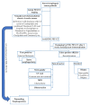
In patients with esophageal cancer undergoing neoadjuvant chemoradiotherapy (nCRT), subsequent restaging with F-18-fluorodeoxyglucose (18F-FDG) positron emission tomography-computed tomography (PET-CT) can reveal the presence of interval metastases, such as liver metastases, in approximately 10% of cases. Nevertheless, it is not uncommon in clinical practice to observe focal FDG uptake in the liver that is not associated with liver metastases but rather with radiation-induced liver injury (RILI), which can result in the overstaging of the disease. Liver radiation damage is also a concern during distal esophageal cancer radiotherapy due to its proximity to the left liver lobe, typically included in the radiation field. Post-CRT, if FDG activity appears in the left or caudate liver lobes, a thorough investigation is needed to confirm or rule out distant metastases. The increased FDG uptake in liver lobes post-CRT often presents a diagnostic dilemma. Distinguishing between radiation-induced liver disease and metastasis is vital for appropriate patient management, necessitating a combination of imaging techniques and an understanding of the factors influencing the radiation response. Diagnosis involves identifying new foci of hepatic FDG avidity on PET/CT scans. Geographic regions of hypoattenuation on CT and well-demarcated regions with specific enhancement patterns on contrast-enhanced CT scans and MRI are characteristic of radiation-induced liver disease (RILD). Lack of mass effect on all three modalities (CT, MRI, PET) indicates RILD. Resolution of abnormalities on subsequent examinations also helps in diagnosing RILD. Moreover, it can also help to rule out occult metastases, thereby excluding those patients from further surgery who will not benefit from esophagectomy with curative intent.
Keywords: F-18-fluorodeoxyglucose (18F-FDG); false liver metastasis; neoadjuvant chemoradiotherapy (nCRT); positron emission tomography–computed tomography (PET-CT); radiation-induced liver disease (RILD); radiation-induced liver injury (RILI).
Conflict of interest statement
The authors declare no conflicts of interest.
Figures


References
-
- Sjoquist K.M., Burmeister B.H., Smithers B.M., Zalcberg J.R., Simes R.J., Barbour A., Gebski V. Survival after neoadjuvant chemotherapy or chemoradiotherapy for resectable oesophageal carcinoma: An updated meta-analysis. Lancet Oncol. 2011;12:681–692. doi: 10.1016/S1470-2045(11)70142-5. - DOI - PubMed
-
- Shapiro J., van Lanschot J.J.B., Hulshof M., van Hagen P., van Berge Henegouwen M.I., Wijnhoven B.P.L., van Laarhoven H.W.M., Nieuwenhuijzen G.A.P., Hospers G.A.P., Bonenkamp J.J., et al. Neoadjuvant chemoradiotherapy plus surgery versus surgery alone for oesophageal or junctional cancer (CROSS): Long-term results of a randomised controlled trial. Lancet Oncol. 2015;16:1090–1098. doi: 10.1016/S1470-2045(15)00040-6. - DOI - PubMed
Publication types
Grants and funding
LinkOut - more resources
Full Text Sources
Research Materials

