Review
. 2024 Feb 28;16(5):984.
doi: 10.3390/cancers16050984.

Impact of Complex Apoptotic Signaling Pathways on Cancer Cell Sensitivity to Therapy

Affiliations
Review

Impact of Complex Apoptotic Signaling Pathways on Cancer Cell Sensitivity to Therapy

Ryungsa Kim et al. Cancers (Basel). .

Abstract

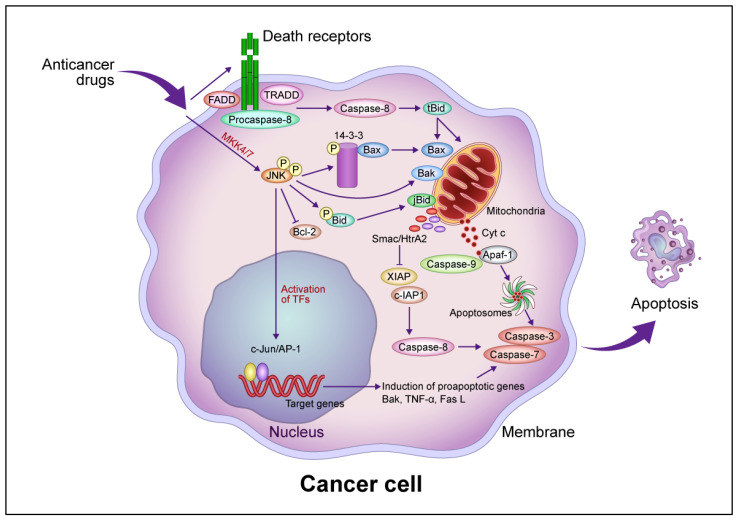
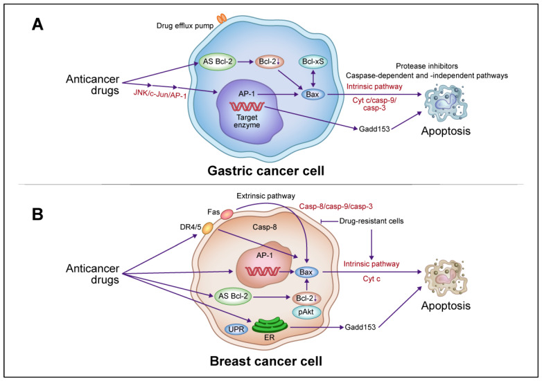
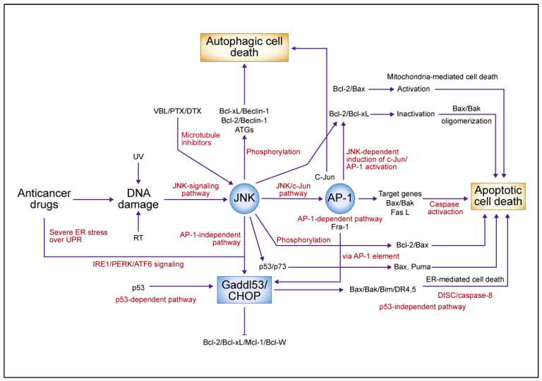
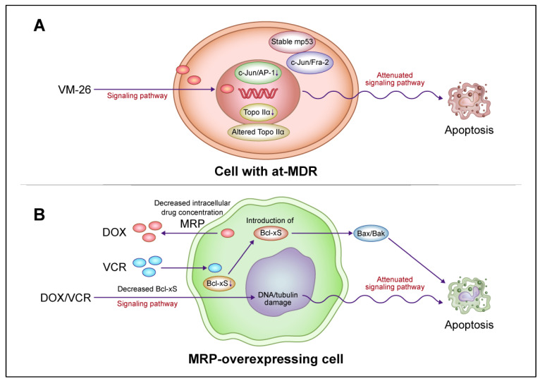
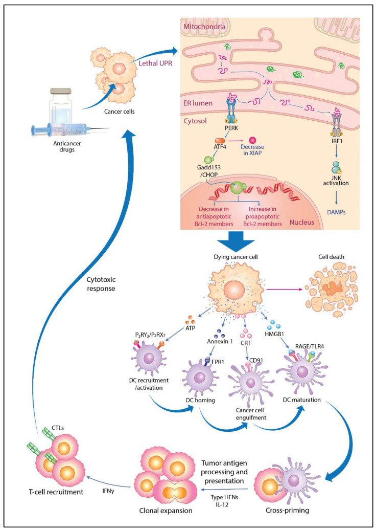
Anticancer drugs induce apoptotic and non-apoptotic cell death in various cancer types. The signaling pathways for anticancer drug-induced apoptotic cell death have been shown to differ between drug-sensitive and drug-resistant cells. In atypical multidrug-resistant leukemia cells, the c-Jun/activator protein 1 (AP-1)/p53 signaling pathway leading to apoptotic death is altered. Cancer cells treated with anticancer drugs undergo c-Jun/AP-1-mediated apoptotic death and are involved in c-Jun N-terminal kinase activation and growth arrest- and DNA damage-inducible gene 153 (Gadd153)/CCAAT/enhancer-binding protein homologous protein pathway induction, regardless of the p53 genotype. Gadd153 induction is associated with mitochondrial membrane permeabilization after anticancer drug treatment and involves a coupled endoplasmic reticulum stress response. The induction of apoptosis by anticancer drugs is mediated by the intrinsic pathway (cytochrome c, Cyt c) and subsequent activation of the caspase cascade via proapoptotic genes (e.g., Bax and Bcl-xS) and their interactions. Anticancer drug-induced apoptosis involves caspase-dependent and caspase-independent pathways and occurs via intrinsic and extrinsic pathways. The targeting of antiapoptotic genes such as Bcl-2 enhances anticancer drug efficacy. The modulation of apoptotic signaling by Bcl-xS transduction increases the sensitivity of multidrug resistance-related protein-overexpressing epidermoid carcinoma cells to anticancer drugs. The significance of autophagy in cancer therapy remains to be elucidated. In this review, we summarize current knowledge of cancer cell death-related signaling pathways and their alterations during anticancer drug treatment and discuss potential strategies to enhance treatment efficacy.

Keywords: anticancer drug; antitumor immunity; cancer cell; cell death; signaling pathway.


Conflict of interest statement

The authors declare that they have no conflicts of interest.

Figures

Figure 1
Figure 1
Common intrinsic and extrinsic apoptotic signaling pathways induced by anticancer drugs. In the intrinsic pathway, anticancer drugs activate JNK via mitogen-activated protein kinase kinase (MKK)4/7, which phosphorylates the 14-3-3 protein, dissociates Bax, and activates Bak. Bax and Bak translocate to the mitochondrial outer membrane and release Cyt c. Cyt c then forms apoptosomes containing apoptotic protease activating factor 1 (Apaf-1) and procaspase-9, activating the caspase cascade that leads to apoptotic cell death. JNK phosphorylates Bid (jBid) and migrates through the mitochondria to release second mitochondria-derived activator of caspases (Smac)/high temperature requirement A2 (HtrA2), which inhibits the X-linked inhibitor of apoptosis protein (XIAP) and cellular inhibitor of apoptosis protein (c-IAP), resulting in the activation of caspase-8 and the caspase cascade. JNK also phosphorylates B-cell lymphoma 2 (Bcl-2) and inhibits its function. In the extrinsic pathway, anticancer drugs activate death receptors that recruit the Fas-associated death domain (FADD), tumor necrosis factor (TNF) receptor-associated death domain (TRADD), and procaspase-8 to form a death-induced signaling complex, which in turn activates caspase-8. Caspase-8 cleaves, generating truncated Bid (tBid), which translocates to the mitochondria, releasing proapoptotic proteins such as Cyt c and Smac/HrtA2. JNK activates c-Jun/activator protein 1 (AP-1), which induces apoptosis-promoting genes such as Bak, TNF-α, and Fas L, in turn activating the caspase cascade leading to apoptotic cell death. Abbreviation: TF, transcription factor. This figure was custom-made by Wiley Editing Services based on our freehand drawing.
Figure 2
Figure 2
Schematic descriptions of the apoptotic death pathways induced by anticancer drugs in gastric and breast cancer cells. (A) In gastric cancer cells, anticancer drugs activate c-Jun N-terminal kinase (JNK)/activator protein 1 (AP-1), induce Bax, release Cyt c, and activate the caspase cascade leading to apoptotic cell death via an intrinsic pathway. AP-1 activation is involved in growth arrest- and DNA damage-inducible gene 153 (Gadd153) induction. Induced Bax and B-cell lymphoma (Bcl)-xS interact to promote this death, which involves caspase-dependent and -independent pathways. Combination therapy with anticancer drugs and antisense (AS) Bcl-2 enhances the therapeutic effect by downregulating Bcl-2 and upregulating Bax, activating a caspase cascade leading to apoptotic cell death. (B) In breast cancer cells, anticancer drugs activate extrinsic pathways via death receptor (DR)4/5 and Fas, leading to the activation of caspase-8 and Bax, release of Cyt c, activation of the caspase cascade, and ultimately apoptotic cell death. In drug-resistant cells, anticancer drugs block extrinsic pathways but activate intrinsic pathways involving the induction of Bax, Cyt c, and the caspase cascade, leading to apoptosis. Combination therapy with anticancer drugs and AS Bcl-2 enhances therapeutic efficacy through the downregulation of Bcl-2 and phosphorylated (p)Akt and upregulation of Bax, which activates the caspase cascade and leads to apoptotic cell death. Abbreviations: Casp, caspase; UPR, unfolded protein response; ER endoplasmic reticulum. This figure was custom-made by Wiley Editing Services based on our freehand drawing.
Figure 3
Figure 3
A summary model for the c-Jun N-terminal kinase (JNK)/c-Jun/activator protein 1 (AP-1)-mediated signaling pathway in anticancer drug-induced cell death. Anticancer drugs that damage DNA and tubulin activate JNK, causing c-Jun/AP-1 to dimerize predominantly with Fra-1 and activate target genes such as Bax/Bak and Fas L, which activate the caspase cascade in an AP-1-dependent pathway leading to apoptotic cell death. JNK phosphorylates and inactivates antiapoptotic proteins such as B-cell lymphoma (Bcl)-2 and Bcl-xL, which in turn activate Bax and cause apoptotic cell death. JNK also activates growth arrest- and DNA damage-inducible gene 153 (Gadd153), which is induced by AP-1-dependent and -independent pathways in endoplasmic reticulum (ER)-mediated cell death. ER stress and p53 induction by anticancer drugs activates Gadd153 and causes ER-mediated cell death. JNK phosphorylates Beclin-1, dissociates from Bcl-2, and induces autophagic cell death. The JNK-mediated upregulation of autophagy-related genes (ATGs) involves the AP-1 transcription factor complex. In response to DNA damage, JNK mediates apoptosis by phosphorylating p53, stabilizing p73, dimerizing p53 and p73, and promoting the expression of proapoptotic target genes such as Bax and Puma. Abbreviations: VBL, vinblastine; PTX, paclitaxel; DTX, docetaxel; UV, ultraviolet; RT, radiation therapy; UPR, unfolded protein response; IRE1, inositol-requiring 1; PERK, protein kinase-like ER kinase; ATF6, activating transcription factor 6; CHOP, CCAAT/enhancer-binding protein homologous protein; DR, death receptor; DISC, death-induced signaling complex; Mcl-1, myeloid cell leukemia 1. This figure was custom-made by Wiley Editing Services based on our freehand drawing.
Figure 4
Figure 4
A model for the interaction of drug resistance-related factors in drug-resistant cancer cells. (A) Altered signaling pathways for apoptotic cell death mediated by c-Jun N-terminal kinase (JNK)/activator protein 1 (AP-1) and mutant p53 (mp53) in lymphoblastic leukemia cells with atypical multidrug resistance (at-MDR). at-MDR is defined by the mutation and reduced expression of topoisomerase IIα (Topo IIα) without drug efflux pump overexpression. Teniposide (VM-26) treatment suppresses c-Jun/AP-1 activation in proportion to the degree of drug resistance. Activated AP-1 consists of a c-Jun/Fra-1 dimer in drug-sensitive cells and c-Jun/Fra-2 and, to a lesser extent, c-Jun/Fra-1 dimers in drug-resistant cells. mp53 induction is more stable and the DNA damage-induced p53 response is attenuated after VM-26 treatment in resistant cells compared with those in sensitive cells. In resistant cells, alterations in apoptotic cell death signaling pathways and target enzymes are regulated independently. (B) Alteration of apoptotic cell death signaling pathways upon the reduction in the proapoptotic protein B-cell lymphoma (Bcl)-xS in ABCC1/multidrug resistance protein (MRP)-overexpressing epidermoid carcinoma cells. The introduction of Bcl-xS promotes apoptotic cell death and partially restores drug sensitivity in resistant cells without affecting the reduction in drug accumulation. The attenuation of the apoptotic cell death pathway via the reduction in Bcl-xS expression and drug accumulation by MRP are regulated independently in resistant cells. Abbreviations: DOX, doxorubicin; VCR, vincristine. This figure was custom-made by Wiley Editing Services based on our freehand drawing.
Figure 5
Figure 5
Molecular mechanisms of immunogenic cell death and tumor-specific immune activation via unfolded protein response (UPR) activation by anticancer drugs. When anticancer drugs are administered, cancer cells exceed the UPR threshold to the point of lethality, activating the double-stranded RNA-activated protein kinase-like ER kinase (PERK) and inositol-requiring 1 (IRE1) signaling pathways and activating transcription factor 4 (ATF4) and growth arrest- and DNA damage-inducible gene 153 (Gadd153)/CCAAT/enhancer-binding protein homologous protein (CHOP) induction, which in turn downregulates X-linked inhibitor of apoptotic protein (XIAP), decreasing antiapoptotic and increasing proapoptotic B-cell lymphoma 2 (Bcl-2) protein expression, leading to cell death. In this process, dying cancer cells externalize calreticulin (CRT) to the membrane surface and release damage-associated molecular patterns (DAMPs) such as ATP, annexin 1, and high mobility group box 1 (HMGB1) for immune activation. Dendritic cells (DCs) are recruited and activated by the secretion of ATP that binds to P2RY2 and P2RX7 receptors. They are then homed by the binding of released annexin 1 to formyl peptide receptor 1 (FPR1). CRT stimulates the phagocytosis of dying cancer cells by binding to CD91 receptors on DCs. HMGB1 release stimulates DC recruitment via binding to receptor for advanced glycation end products (RAGE) receptors and induces DC maturation via toll-like receptor 4 (TLR4) signaling. Mature DCs migrate to lymph nodes, where they cross-prime and cause the clonal expansion of T cells, including interleukin 12 (IL-12) and type I interferon (IFN). IFN-γ-producing T cells are recruited and exert cytotoxic responses to eradicate cancer cells. Tumor antigens (TAs) released from dying cells are taken up by immature DCs, which then activate and mature. TAs and tumor-associated antigens (TAAs) are processed in mature DCs and presented to CD8+ T cells as major histocompatibility complex I (MHC I) molecules, generating cytotoxic T lymphocytes (CTLs) for tumor-specific immune responses. This figure was custom-made by Wiley Editing Services based on our freehand drawing.
Figure 6
Figure 6
The enhancement of drug sensitivity via molecular therapies targeting tumor growth and antitumor immunity. Human epidermal growth factor receptor (HER) 2- and HER3-targeted antibody–drug conjugate (ADC) products, including topoisomerase I (Topo I) inhibitors such as trastuzumab deluxecan (T-DXd) and patritumab deluxecan (HER3-DXd), have considerable antitumor effects in patients who have undergone HER2-targeted therapy. These ADCs are internalized into cancer cells and release Topo I inhibitors that are not cross-resistant to anthracyclines and taxanes, leading to cell death. The released payload penetrates adjacent cancer cells through a bystander effect. The ADC sacituzumab govitecan targets trophoblast antigen 2 (Trop-2), a surface protein bound to the Topo I inhibitor SN-38, which is released intracellularly and extracellularly. Cell cycle inhibitors targeting cyclin-dependent kinase 4/6 (CDK4/6) in the G1–S phase transition have shown promise when used in combination with endocrine therapy for patients with hormone receptor (HR)-positive HER2-negative breast cancer. The development of cancer stem cells (CSCs) with persisting drug resistance is a major obstacle in chemotherapy. CSC-targeting therapies, which target several signaling pathways involved in cancer survival, may be employed effectively in combination with conventional therapy to eradicate residual tumor cells for the curing of cancer. The combined use of anticancer drugs and immune checkpoint inhibitors, such as anti-programmed cell death 1 (PD-1), anti-programed cell death ligand 1 (PD-L1), and anti-cytotoxic T lymphocyte antigen-4 (CTLA-4) antibodies, enhances therapeutic efficacy by increasing antitumor immune activity. Abbreviations: MHC, major histocompatibility complex; TCR, T-cell receptor; APC, antigen-presenting cell. This figure was custom-made by Wiley Editing Services based on our freehand drawing.

Similar articles

Cited by

References

    1. Hanahan D., Weinberg R.A. Hallmarks of cancer: The next generation. Cell. 2011;144:646–674. doi: 10.1016/j.cell.2011.02.013. - DOI - PubMed
    1. Hotchkiss R.S., Strasser A., McDunn J.E., Swanson P.E. Cell death. N. Engl. J. Med. 2009;361:1570–1583. doi: 10.1056/NEJMra0901217. - DOI - PMC - PubMed
    1. Baig S., Seevasant I., Mohamad J., Mukheem A., Huri H.Z., Kamarul T. Potential of apoptotic pathway-targeted cancer therapeutic research: Where do we stand? Cell Death Dis. 2016;7:e2058. doi: 10.1038/cddis.2015.275. - DOI - PMC - PubMed
    1. Plati J., Bucur O., Khosravi-Far R. Apoptotic cell signaling in cancer progression and therapy. Integr. Biol. 2011;3:279–296. doi: 10.1039/c0ib00144a. - DOI - PMC - PubMed
    1. Kroemer G., Galluzzi L., Brenner C. Mitochondrial membrane permeabilization in cell death. Physiol. Rev. 2007;87:99–163. doi: 10.1152/physrev.00013.2006. - DOI - PubMed