Review
. 2024 Feb 29;16(5):991.
doi: 10.3390/cancers16050991.

Epigenetic Mechanisms in Latent Epstein-Barr Virus Infection and Associated Cancers

Affiliations
Review

Epigenetic Mechanisms in Latent Epstein-Barr Virus Infection and Associated Cancers

Atharva S Torne et al. Cancers (Basel). .

Abstract

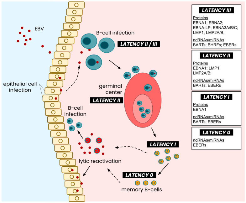
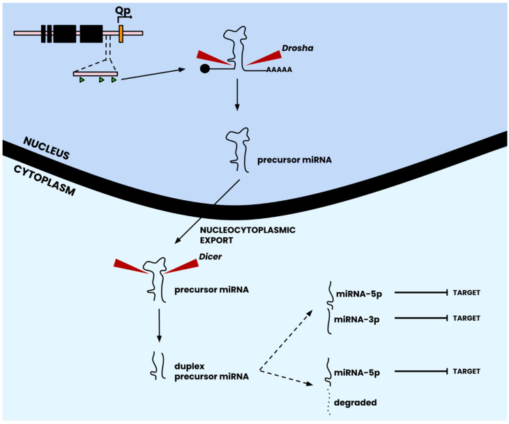
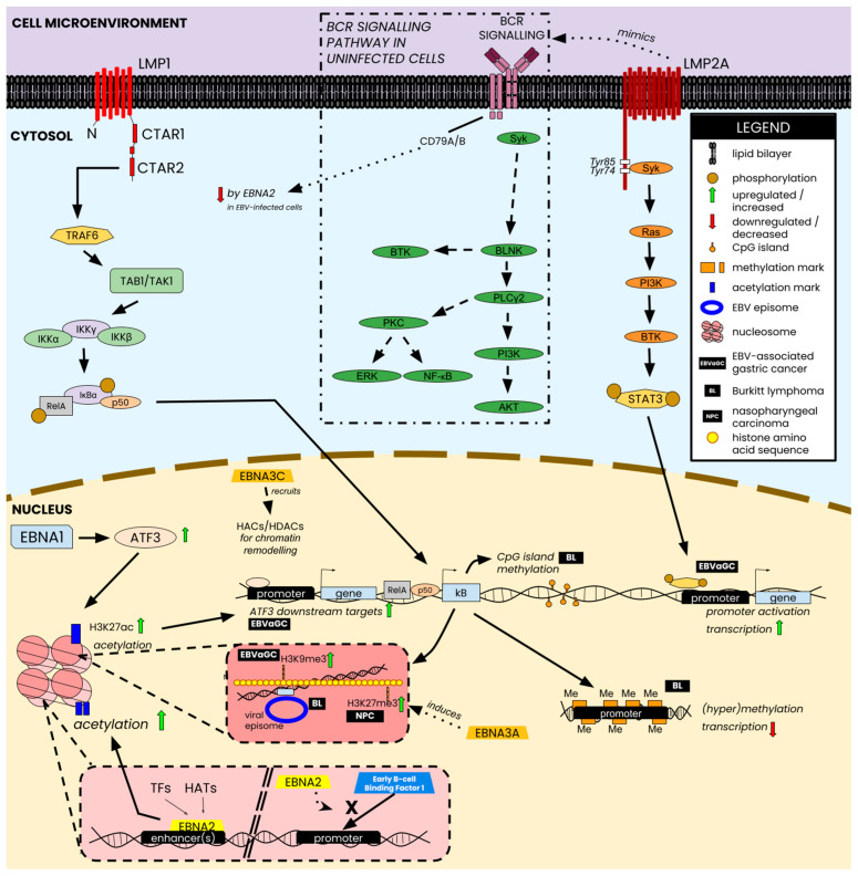
The Epstein-Barr Virus (EBV) is a double-stranded DNA-based human tumor virus that was first isolated in 1964 from lymphoma biopsies. Since its initial discovery, EBV has been identified as a major contributor to numerous cancers and chronic autoimmune disorders. The virus is particularly efficient at infecting B-cells but can also infect epithelial cells, utilizing an array of epigenetic strategies to establish long-term latent infection. The association with histone modifications, alteration of DNA methylation patterns in host and viral genomes, and microRNA targeting of host cell factors are core epigenetic strategies that drive interactions between host and virus, which are necessary for viral persistence and progression of EBV-associated diseases. Therefore, understanding epigenetic regulation and its role in post-entry viral dynamics is an elusive area of EBV research. Here, we present current outlooks of EBV epigenetic regulation as it pertains to viral interactions with its host during latent infection and its propensity to induce tumorigenesis. We review the important epigenetic regulators of EBV latency and explore how the strategies involved during latent infection drive differential epigenetic profiles and host-virus interactions in EBV-associated cancers.

Keywords: EBV-associated cancers; Epstein–Barr virus; host virus interactions; microRNAs; tumor viruses; viral epigenetics.


Conflict of interest statement

The authors declare no conflicts of interest.

Figures

Figure 1
Figure 1
Linearized structure of the wildtype EBV latent genome. The wildtype genome is composed of four latent promoters (Cp, red; Wp, magenta; Qp, orange; LMPp, yellow) that produce mRNA transcripts for the eight major latent antigens. The effect of dysregulated latent promoter activity can sometimes be mitigated by redirecting some latent transcription to certain lytic promoters, such as Fp (light blue) [20]. The major antigens are indicated above as black bars that show the relative position of each antigen’s coding exons. The LMP coding region traverses the terminal repeats and loops back into the ‘start’ of this linear structure. Non-coding RNAs (ncRNAs; termed EBERs in EBV and shown as green triangles) are encoded in a ~700 bp fragment preceding the origin of replication (OriP). Each major latent protein coding region is separated by regions encoding microRNAs (miRNAs); the BamHI rightward fragment 1 (BHRF1) and BamHI rightward A transcript (BART) regions encode twenty-five genomic loci (green triangles; indicative of location only) that produce approximately forty-four mature miRNAs. Spontaneous deletions in the BART region (red underline) are deleted in the B95-8 laboratory strain. Figure adapted from [21].
Figure 2
Figure 2
Latency types across the EBV life cycle. Following the entry and infection of host B-cells, EBV undergoes latency III expression, which eventually degrades into latency II expression of its latent genome. As infected cells enter the germinal center and undergo maturation, the EBV genome is suppressed into latency II expression of EBERs, LMPs, and EBNA1, which further devolves into latency I expression as mature B-cells exit the germinal center. Fully mature memory B-cells exhibit a near-quiescent EBV latent genome, where only EBERs are expressed in minimal quantities. Lytic reactivation of the virus enables viral capsid replication and further infection of cells, both lymphocytic and epithelial. Figure adapted from [27].
Figure 3
Figure 3
Canonical pathway of miRNA biogenesis. Following transcription from the viral genome, precursor miRNA is processed by the Drosha complex to remove the 5′ cap and poly-A tail to yield a precursor miRNA transcript in its classical stem-loop structure. Following nucleocytoplasmic export, the precursor miRNA transcript is acted upon by the Dicer complex, which removes the stem-loop sequence to yield a duplexed precursor miRNA transcript. The duplex dissociates into two strands, one of which is degraded and one that produces a unique mature miRNA. Depending on the miRNA locus, however, the duplex precursor can also dissociate into two unique, matured miRNAs (which is the case for many EBV miRNAs; see Table 2). Mature miRNAs subsequently are localized to their area of function for targeting. Figure adapted from [82].
Figure 4
Figure 4
Selected epigenetic changes and host-virus interactions perpetrated by EBV nuclear antigens and latent membrane proteins. Nuclear antigens (e.g., EBNA1) interact directly with transcription factors and/or intermediate proteins of signaling pathways that lead to the deposition of acetylation marks on histones, which translate into the upregulation of downstream targets of the corresponding intermediate, shown in the figure, as ATF3. EBNA2 can also bind DNA sites in the enhancer and promote transcription by attracting histone acetyltransferases (HATs) or transcription factors (TFs) to those sites. Alternatively, EBNA2 can also block the binding of transcription factors to promoters and repress the downstream targets of TFs, such as the EBNA2-mediated repression of CD79B via TF-blocking at the promoter. From a chromatin modeling perspective, EBNA3C recruits histone HATs and histone deacetylases (HDACs) to controlhistone configuration, thereby controlling transcriptional access for both viral and host TFs. Membrane proteins perpetrate similar changes via modulation of signaling activity. LMP1 modulates the NF-kB signaling pathway to inflict epigenetic changes such as CpG island methylation, lysine-specific histone methylation, and promoter-specific methylation to control gene transcription in EBVaGC, BL, and NPC. LMP2A mimics the BCR signaling cascade and directly targets the phosphorylation of STAT3, which subsequently binds and activates key promoters such as those that controls transcription of some factors EBVaGC. Figure composed using information from [28,38,179,180,181,182,183].

Similar articles

Cited by

References

    1. Rous P., Murphy J.B. On The Causation by Filterable Agents of Three Distinct Chicken Tumors. J. Exp. Med. 1914;19:52–68. doi: 10.1084/jem.19.1.52. - DOI - PMC - PubMed
    1. Rous P., Murphy J.B., Tytler W.H. A filterable agent the cause of a second chicken-tumor, an osteochondrosarcoma. J. Am. Med. Assoc. 1912;59:1793–1794. doi: 10.1001/jama.1912.04270110207011. - DOI
    1. Burkitt D. A sarcoma involving the jaws in African children. Br. J. Surg. 1958;46:218–223. doi: 10.1002/bjs.18004619704. - DOI - PubMed
    1. Epstein M.A., Achong B.G., Barr Y.M. Virus particles in cultured lymphoblasts from burkitt’s lymphoma. Lancet. 1964;1:702–703. doi: 10.1016/S0140-6736(64)91524-7. - DOI - PubMed
    1. Saha A., Kaul R., Murakami M., Robertson E.S. Tumor viruses and cancer biology. Cancer Biol. Ther. 2010;10:961–978. doi: 10.4161/cbt.10.10.13923. - DOI - PMC - PubMed

LinkOut - more resources