

The NF1+/- Immune Microenvironment: Dueling Roles in Neurofibroma Development and Malignant Transformation
- PMID: 38473354
- PMCID: PMC10930863
- DOI: 10.3390/cancers16050994
The NF1+/- Immune Microenvironment: Dueling Roles in Neurofibroma Development and Malignant Transformation
Abstract
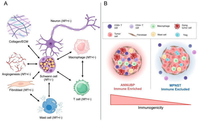
Neurofibromatosis type 1 (NF1) is a common genetic disorder resulting in the development of both benign and malignant tumors of the peripheral nervous system. NF1 is caused by germline pathogenic variants or deletions of the NF1 tumor suppressor gene, which encodes the protein neurofibromin that functions as negative regulator of p21 RAS. Loss of NF1 heterozygosity in Schwann cells (SCs), the cells of origin for these nerve sheath-derived tumors, leads to the formation of plexiform neurofibromas (PNF)-benign yet complex neoplasms involving multiple nerve fascicles and comprised of a myriad of infiltrating stromal and immune cells. PNF development and progression are shaped by dynamic interactions between SCs and immune cells, including mast cells, macrophages, and T cells. In this review, we explore the current state of the field and critical knowledge gaps regarding the role of NF1(Nf1) haploinsufficiency on immune cell function, as well as the putative impact of Schwann cell lineage states on immune cell recruitment and function within the tumor field. Furthermore, we review emerging evidence suggesting a dueling role of Nf1+/- immune cells along the neurofibroma to MPNST continuum, on one hand propitiating PNF initiation, while on the other, potentially impeding the malignant transformation of plexiform and atypical neurofibroma precursor lesions. Finally, we underscore the potential implications of these discoveries and advocate for further research directed at illuminating the contributions of various immune cells subsets in discrete stages of tumor initiation, progression, and malignant transformation to facilitate the discovery and translation of innovative diagnostic and therapeutic approaches to transform risk-adapted care.
Keywords: Schwann cells; T cells; atypical neurofibroma; biomarkers; immune microenvironment; immunotherapy; macrophages; malignant peripheral nerve sheath tumor; mast cells; neurofibromatosis type 1 (NF1); plexiform neurofibroma.
Conflict of interest statement
The authors declare no conflicts of interest.
Figures

Similar articles
-
Dual mTORC1/2 inhibition induces anti-proliferative effect in NF1-associated plexiform neurofibroma and malignant peripheral nerve sheath tumor cells.Oncotarget. 2016 Jun 14;7(24):35753-35767. doi: 10.18632/oncotarget.7099. Oncotarget. 2016. PMID: 26840085 Free PMC article.
-
After Nf1 loss in Schwann cells, inflammation drives neurofibroma formation.Neurooncol Adv. 2019 Nov 22;2(Suppl 1):i23-i32. doi: 10.1093/noajnl/vdz045. eCollection 2020 Jul. Neurooncol Adv. 2019. PMID: 32642730 Free PMC article. Review.
-
Spatial Gene-Expression Profiling Unveils Immuno-oncogenic Programs of NF1-Associated Peripheral Nerve Sheath Tumor Progression.Clin Cancer Res. 2024 Mar 1;30(5):1038-1053. doi: 10.1158/1078-0432.CCR-23-2548. Clin Cancer Res. 2024. PMID: 38127282 Free PMC article.
-
Cdkn2a Loss in a Model of Neurofibroma Demonstrates Stepwise Tumor Progression to Atypical Neurofibroma and MPNST.Cancer Res. 2020 Nov 1;80(21):4720-4730. doi: 10.1158/0008-5472.CAN-19-1429. Epub 2020 Aug 19. Cancer Res. 2020. PMID: 32816910 Free PMC article.
-
Modeling tumors of the peripheral nervous system associated with Neurofibromatosis type 1: Reprogramming plexiform neurofibroma cells.Stem Cell Res. 2020 Dec;49:102068. doi: 10.1016/j.scr.2020.102068. Epub 2020 Oct 29. Stem Cell Res. 2020. PMID: 33160273 Review.
Cited by
-
UBR5 in Tumor Biology: Exploring Mechanisms of Immune Regulation and Possible Therapeutic Implications in MPNST.Cancers (Basel). 2025 Jan 7;17(2):161. doi: 10.3390/cancers17020161. Cancers (Basel). 2025. PMID: 39857943 Free PMC article. Review.
-
Granulocyte-Macrophage Colony Stimulating Factor Receptor Contributes to Plexiform Neurofibroma Initiation.Cancers (Basel). 2025 Mar 6;17(5):905. doi: 10.3390/cancers17050905. Cancers (Basel). 2025. PMID: 40075752 Free PMC article.
-
DLK1 Distinguishes Subsets of NF1-Associated Malignant Peripheral Nerve Sheath Tumors with Divergent Molecular Signatures.Clin Cancer Res. 2025 May 15;31(10):1988-2009. doi: 10.1158/1078-0432.CCR-24-3029. Clin Cancer Res. 2025. PMID: 40063513
-
A Case Report and Review of Diagnostic and Therapeutic Challenges of a Malignant Peripheral Nerve Sheath Tumor in the Foot.Cureus. 2024 Jul 29;16(7):e65669. doi: 10.7759/cureus.65669. eCollection 2024 Jul. Cureus. 2024. PMID: 39205702 Free PMC article.
References
Publication types
Grants and funding
LinkOut - more resources
Full Text Sources
Molecular Biology Databases
Research Materials
Miscellaneous
