. 2024 Mar 4;16(5):1046.
doi: 10.3390/cancers16051046.

Haploinsufficiency of Adenomatous Polyposis Coli Coupled with Kirsten Rat Sarcoma Viral Oncogene Homologue Activation and P53 Loss Provokes High-Grade Glioblastoma Formation in Mice

Affiliations

Haploinsufficiency of Adenomatous Polyposis Coli Coupled with Kirsten Rat Sarcoma Viral Oncogene Homologue Activation and P53 Loss Provokes High-Grade Glioblastoma Formation in Mice

Kuan-Te Fang et al. Cancers (Basel). .

Abstract

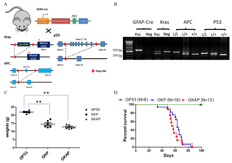
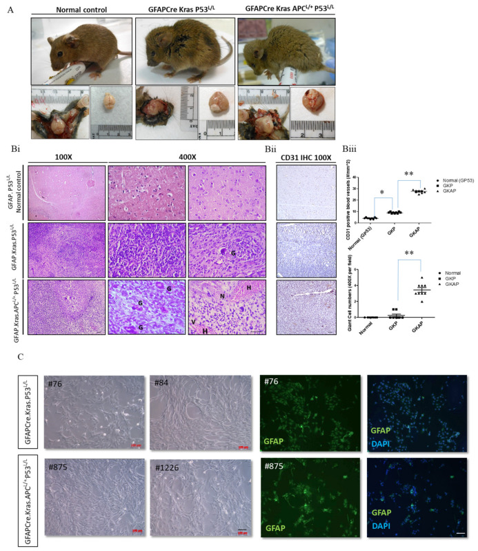
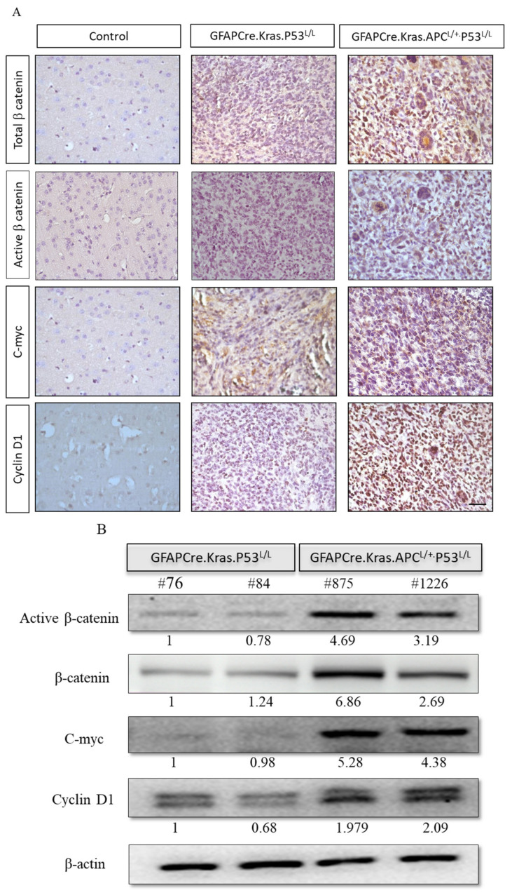
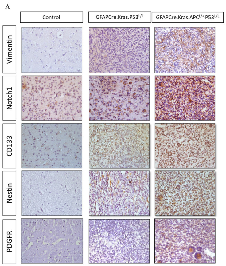
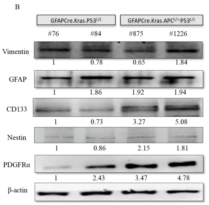
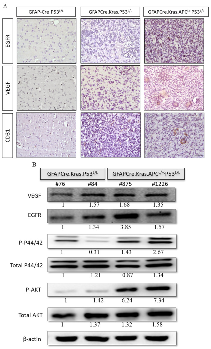
Glioblastoma multiforme (GBM) is the most common and deadly type of brain tumor originating from glial cells. Despite decades of clinical trials and research, there has been limited success in improving survival rates. However, molecular pathology studies have provided a detailed understanding of the genetic alterations associated with the formation and progression of glioblastoma-such as Kirsten rat sarcoma viral oncogene homolog (KRAS) signaling activation (5%), P53 mutations (25%), and adenomatous polyposis coli (APC) alterations (2%)-laying the groundwork for further investigation into the biological and biochemical basis of this malignancy. These analyses have been crucial in revealing the sequential appearance of specific genetic lesions at distinct histopathological stages during the development of GBM. To further explore the pathogenesis and progression of glioblastoma, here, we developed the glial-fibrillary-acidic-protein (GFAP)-Cre-driven mouse model and demonstrated that activated KRAS and p53 deficiencies play distinct and cooperative roles in initiating glioma tumorigenesis. Additionally, the combination of APC haploinsufficiency with mutant Kras activation and p53 deletion resulted in the rapid progression of GBM, characterized by perivascular inflammation, large necrotic areas, and multinucleated giant cells. Consequently, our GBM models have proven to be invaluable resources for identifying early disease biomarkers in glioblastoma, as they closely mimic the human disease. The insights gained from these models may pave the way for potential advancements in the diagnosis and treatment of this challenging brain tumor.

Keywords: APC haploinsufficiency; animal models; giant cells; glioblastoma multiforme (GBM).


Conflict of interest statement

The authors declare no conflicts of interest.

Figures

Figure 1
Figure 1
Targeting mutant Kras activation with APC and p53 deletion to induce GBM formation in mice. (A) Schematic diagram for the genetically modified GBM mouse mating setup. (B) Specifically genotyping PCR was conducted for the genotyping of animals and the detection of the LoxP product of each targeted allele, with PCR products visualized via agarose gel electrophoresis and displaying the expected sizes: GFAP-Cre (600 bp), Kras size (LoxP: 327 bp; wild type: 450 bp), APC (LoxP: 430 bp; wild type: 320 bp) and P53 (LoxP: 370 bp; wildtype: 288 bp). Weight curve (C) and survival (D) alterations for GFAP-Cre; p53L/L (GP53 serves the normal control group), GFAP-Cre; Kras; p53L/L (GKP) and GFAP-Cre; Kras; APCL/+; p53L/L (GKAP) mice. ** p < 0.01.
Figure 2
Figure 2
Pathohistological analyses showing glioblastoma formation in GFAP-Cre; KrasG12D; P53L/L and GFAP-Cre; KrasG12D; APCL/+; P53L/L mice. (A) Gross anatomy analysis of GFAP-Cre; P53L/L (GP53 normal control), GFAP-Cre; KrasG12D; P53L/L (GKP), and GFAP-Cre; KrasG12D; APCL/+; P53L/L (GKAP) mice. (B) (i) Representative hematoxylin-and-eosin (H&E)-stained brain sections (the temporal lobes) representing different indicated genotypes; GFAP-Cre; P53L/L control; GFAP-Cre; KrasG12D; P53L/L and GFAP-Cre; KrasG12D; APCL/+; P53L/L mice. Key features include multinucleated giant cells (G), hemorrhage (H), increased cellularity, vascularity (V), and necrosis (N). Bars: 100 μm. (ii) IHC staining of CD31 on the brain sections derived from GFAP-Cre; P53L/L, GFAP-Cre; KrasG12D; P53L/L, and GFAP-Cre; KrasG12D; APCL/+; P53L/L mice. (iii) The numbers of CD31-labeled blood vessels and giant cells were counted in six non-overlapping microscopic fields of the indicated genotypes. * p < 0.05; ** p < 0.01. (C) Primary cell cultures derived from the brains of GFAP-Cre; KrasG12D; P53L/L, and GFAP-Cre; KrasG12D; APCL/+; P53L/L mice. Morphological characteristics of GFAP-Cre; KrasG12D; P53L/L (#76; #84), and GFAP-Cre; KrasG12D; APCL/+; P53L/L (#875; #1226) cells are presented, respectively. Murine GBM primary cell lines, confirmed as glioblastoma, displayed GFAP glial marker immunopositivity (FITC) with DAPI-stained blue nuclei. Bars: 100 μm.
Figure 3
Figure 3
Analysis of proliferative and apoptotic activities in murine GBM. (Ai) TUNEL assays were conducted to compare the extent of necrosis in GFAP-Cre; KrasG12D; APCL/+; P53L/L (GKAP) brain sections with the indicated genotypes. Cell proliferation was evaluated through whole-mount immunohistochemistry using anti-phosphohistone H3 and anti-Ki-67 antibodies, as indicated. Bars: 100 μm. (Aii) The percentage of TUNEL-, Ki67-, and pHistone3-positive cells were quantified by ImageJ software ver. 1.45. ** p < 0.01. (B) Proliferative rates were determined via MTT assays on primary cells cultured from the brains of GFAP-Cre; KrasG12D; P53L/L and GFAP-Cre; KrasG12D; APCL/+; P53L/L mice. (C) The colony formation assays demonstrated the impact of APC haploinsufficiency on the ability of murine primary GBM cells to form colonies.
Figure 4
Figure 4
Enhanced activation of the Wnt/β-Catenin pathway in GBM from GFAP-Cre; KrasG12D; APCL/+; P53L/L mice. (A) Immunohistochemistry analysis reveals the activation of the Wnt/β-catenin pathway, as evidenced by the high expression of β-catenin, c-myc, and cyclin D1 proteins. Bars: 100 μm. (B) Western blot analysis shows the expression of active β-catenin, β-catenin, c-myc, and cyclin D1 in primary GBM cells isolated from GFAP-Cre; KrasG12D; P53L/L (#76; #84) and GFAP-Cre; KrasG12D; APCL/+; P53L/L (#875; #1226) mice. β-Actin levels were used for protein loading normalization. Relative pixel intensities were measured via densitometry analysis using ImageJ 1.45s software.
Figure 5
Figure 5
Regulation of cancer-stem-like properties in GBM mice with APC haploinsufficiency. (A) Immunohistochemistry analysis was performed to evaluate the expression levels of Vimentin, Notch1, CD133, Nestin, and PDGFRα proteins in formalin-fixed brain tissues obtained from GFAP-Cre; P53L/L, GFAP-Cre; KrasG12D; P53L/L, and GFAP-Cre; KrasG12D; APCL/+; P53L/L mice. Bars: 100 μm. (B) Western blot analysis was conducted to detect the protein levels of Vimentin, GFAP, CD133, Nestin, and PDGFRα in primary GBM cells derived from GFAP-Cre; KrasG12D; P53L/L (#76; #84) and GFAP-Cre; KrasG12D; APCL/+; P53L/L (#875; #1226) mice, respectively. The expression of β-actin was used to ensure uniform protein loading across all lanes. Relative pixel intensities were measured via densitometry analysis using ImageJ 1.45s software.
Figure 5
Figure 5
Regulation of cancer-stem-like properties in GBM mice with APC haploinsufficiency. (A) Immunohistochemistry analysis was performed to evaluate the expression levels of Vimentin, Notch1, CD133, Nestin, and PDGFRα proteins in formalin-fixed brain tissues obtained from GFAP-Cre; P53L/L, GFAP-Cre; KrasG12D; P53L/L, and GFAP-Cre; KrasG12D; APCL/+; P53L/L mice. Bars: 100 μm. (B) Western blot analysis was conducted to detect the protein levels of Vimentin, GFAP, CD133, Nestin, and PDGFRα in primary GBM cells derived from GFAP-Cre; KrasG12D; P53L/L (#76; #84) and GFAP-Cre; KrasG12D; APCL/+; P53L/L (#875; #1226) mice, respectively. The expression of β-actin was used to ensure uniform protein loading across all lanes. Relative pixel intensities were measured via densitometry analysis using ImageJ 1.45s software.
Figure 6
Figure 6
Levels of expression of the VEGF and EGFR kinase pathways in GBM derived from GFAP-Cre; P53L/L, GFAP-Cre; KrasG12D; P53L/L, and GFAP-Cre; KrasG12D; APCL/+; P53L/L mice. (A) Immunohistochemical analysis of EGFR and VEGF in brain tissue sections from GFAP-Cre; P53L/L, GFAP-Cre; KrasG12D; P53L/L, and GFAP-Cre; KrasG12D; APCL/+; P53L/L mouse brain tissues. Bars: 100 μm. (B) Western blot analyses assess the expression levels of EGFR, VEGF, total downstream markers p44/42, P-p44/42, P-Akt, and total Akt protein in GFAP-Cre; KrasG12D; P53L/L (#76; #84) and GFAP-Cre; KrasG12D; APCL/+; P53L/L (#875; #1226) cells. β-Actin was used as a loading control. Relative pixel intensities were measured via densitometry analysis using ImageJ 1.45s software.

Similar articles

References

    1. Van den Bent M.J., Geurts M., French P.J., Smits M., Capper D., Bromberg J.E.C., Chang S.M. Primary brain tumours in adults. Lancet. 2023;402:1564–1579. doi: 10.1016/S0140-6736(23)01054-1. - DOI - PubMed
    1. Jones T.S., Holland E.C. Molecular pathogenesis of malignant glial tumors. Toxicol. Pathol. 2011;39:158–166. doi: 10.1177/0192623310387617. - DOI - PubMed
    1. Krex D., Klink B., Hartmann C., von Deimling A., Piestch T., Simon M., Sabel M., Steinbacj J.P., Heese O., Reifenberger G., et al. Long-term survival with glioblastoma multiforme. Pt 10Brain. 2007;130:2596–2606. doi: 10.1093/brain/awm204. - DOI - PubMed
    1. Ohgaki H., Kleihues P. Population-based studies on incidence, survival rates, and genetic alterations in astrocytic and oligodendroglial gliomas. J. Neuropathol. Exp. Neurol. 2005;64:479–489. doi: 10.1093/jnen/64.6.479. - DOI - PubMed
    1. Smoll N.R., Schaller K., Gautschi O.P. Long-term survival of patients with glioblastoma multiforme (GBM) J. Clin. Neurosci. 2013;20:670–675. doi: 10.1016/j.jocn.2012.05.040. - DOI - PubMed

LinkOut - more resources