

Use of microRNAs as Diagnostic, Prognostic, and Therapeutic Tools for Glioblastoma
- PMID: 38473710
- PMCID: PMC10931459
- DOI: 10.3390/ijms25052464
Use of microRNAs as Diagnostic, Prognostic, and Therapeutic Tools for Glioblastoma
Abstract
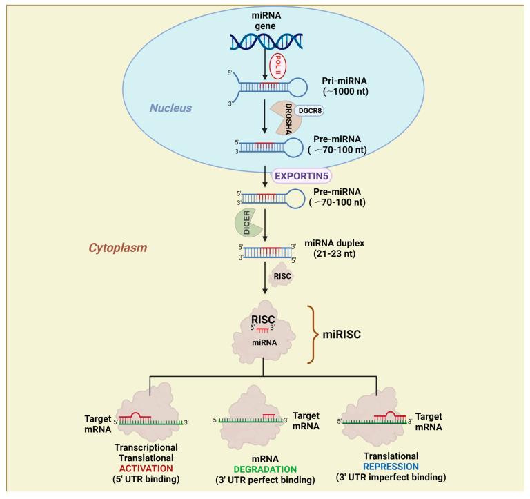
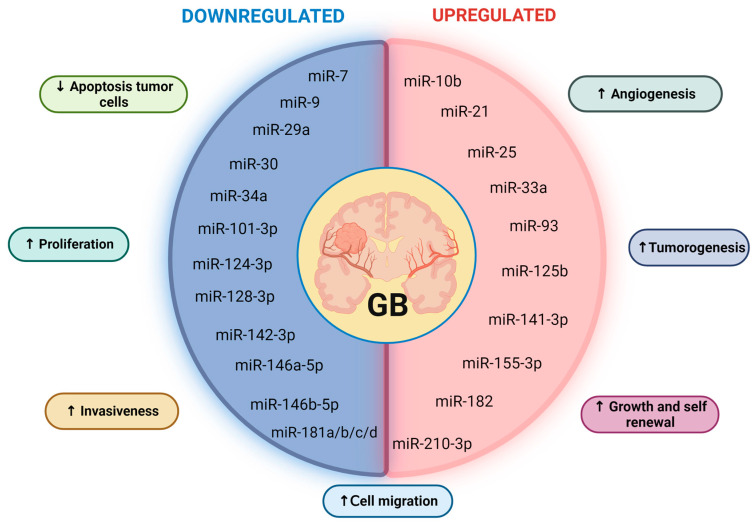
Glioblastoma (GB) is the most aggressive and common type of cancer within the central nervous system (CNS). Despite the vast knowledge of its physiopathology and histology, its etiology at the molecular level has not been completely understood. Thus, attaining a cure has not been possible yet and it remains one of the deadliest types of cancer. Usually, GB is diagnosed when some symptoms have already been presented by the patient. This diagnosis is commonly based on a physical exam and imaging studies, such as computed tomography (CT) and magnetic resonance imaging (MRI), together with or followed by a surgical biopsy. As these diagnostic procedures are very invasive and often result only in the confirmation of GB presence, it is necessary to develop less invasive diagnostic and prognostic tools that lead to earlier treatment to increase GB patients' quality of life. Therefore, blood-based biomarkers (BBBs) represent excellent candidates in this context. microRNAs (miRNAs) are small, non-coding RNAs that have been demonstrated to be very stable in almost all body fluids, including saliva, serum, plasma, urine, cerebrospinal fluid (CFS), semen, and breast milk. In addition, serum-circulating and exosome-contained miRNAs have been successfully used to better classify subtypes of cancer at the molecular level and make better choices regarding the best treatment for specific cases. Moreover, as miRNAs regulate multiple target genes and can also act as tumor suppressors and oncogenes, they are involved in the appearance, progression, and even chemoresistance of most tumors. Thus, in this review, we discuss how dysregulated miRNAs in GB can be used as early diagnosis and prognosis biomarkers as well as molecular markers to subclassify GB cases and provide more personalized treatments, which may have a better response against GB. In addition, we discuss the therapeutic potential of miRNAs, the current challenges to their clinical application, and future directions in the field.
Keywords: GBM; RNA therapy; glioblastoma; glioblastoma multiforme; glioma; glioma treatment; miRNAs; microRNAs; non-coding RNAs.
Conflict of interest statement
The authors declare no conflicts of interest.
Figures
Similar articles
-
Analysis of Glioblastoma Patients' Plasma Revealed the Presence of MicroRNAs with a Prognostic Impact on Survival and Those of Viral Origin.PLoS One. 2015 May 7;10(5):e0125791. doi: 10.1371/journal.pone.0125791. eCollection 2015. PLoS One. 2015. PMID: 25950799 Free PMC article.
-
Profiling of novel circulating microRNAs as a non-invasive biomarker in diagnosis and follow-up of high and low-grade gliomas.Clin Neurol Neurosurg. 2020 Mar;190:105652. doi: 10.1016/j.clineuro.2019.105652. Epub 2019 Dec 27. Clin Neurol Neurosurg. 2020. PMID: 31896490
-
MicroRNA based theranostics for brain cancer: basic principles.J Exp Clin Cancer Res. 2019 May 29;38(1):231. doi: 10.1186/s13046-019-1180-5. J Exp Clin Cancer Res. 2019. PMID: 31142339 Free PMC article. Review.
-
Systematic characterization and biological functions of non-coding RNAs in glioblastoma.Cell Prolif. 2023 Mar;56(3):e13375. doi: 10.1111/cpr.13375. Epub 2022 Dec 1. Cell Prolif. 2023. PMID: 36457281 Free PMC article. Review.
-
Angioregulatory role of miRNAs and exosomal miRNAs in glioblastoma pathogenesis.Biomed Pharmacother. 2022 Apr;148:112760. doi: 10.1016/j.biopha.2022.112760. Epub 2022 Feb 25. Biomed Pharmacother. 2022. PMID: 35228062 Review.
Cited by
-
Inhibition of circular JUN prevents the proliferation and invasion of glioblastoma via miR-3064-IGFBP5 axis.J Cell Mol Med. 2024 Sep;28(18):e70098. doi: 10.1111/jcmm.70098. J Cell Mol Med. 2024. PMID: 39307884 Free PMC article.
-
Harnessing the Role of ESR1 in Breast Cancer: Correlation with microRNA, lncRNA, and Methylation.Int J Mol Sci. 2025 Mar 27;26(7):3101. doi: 10.3390/ijms26073101. Int J Mol Sci. 2025. PMID: 40243758 Free PMC article.
-
Regulation of autophagy by non-coding RNAs in human glioblastoma.Med Oncol. 2024 Oct 7;41(11):260. doi: 10.1007/s12032-024-02513-3. Med Oncol. 2024. PMID: 39375229 Review.
-
Identification of Deregulated miRNAs and mRNAs Involved in Tumorigenesis and Detection of Glioblastoma Patients Applying Next-Generation RNA Sequencing.Pharmaceuticals (Basel). 2025 Mar 19;18(3):431. doi: 10.3390/ph18030431. Pharmaceuticals (Basel). 2025. PMID: 40143207 Free PMC article.
-
Exosomes in Regulating miRNAs for Biomarkers of Neurodegenerative Disorders.Mol Neurobiol. 2025 Jun;62(6):7576-7596. doi: 10.1007/s12035-025-04733-8. Epub 2025 Feb 7. Mol Neurobiol. 2025. PMID: 39918711 Review.
References
Publication types
MeSH terms
Substances
LinkOut - more resources
Full Text Sources
Research Materials
