. 2024 Feb 20;25(5):2468.
doi: 10.3390/ijms25052468.

Knockdown of DNMT1 Induces SLCO3A1 to Promote Follicular Growth by Enhancing the Proliferation of Granulosa Cells in Mammals

Affiliations

Knockdown of DNMT1 Induces SLCO3A1 to Promote Follicular Growth by Enhancing the Proliferation of Granulosa Cells in Mammals

Shuo Li et al. Int J Mol Sci. .

Abstract

In female mammals, the proliferation and apoptosis of granulosa cells (GCs) have been shown to determine the fate of follicles. DNA methyltransferases (DNMTs) and SLCO3A1 have been reported to be involved in the survival of GCs and follicular growth. However, the molecular mechanisms enabling DNMTs to regulate the expression of SLCO3A1 to participate in follicular growth are unclear. In this study, we found that the knockdown of DNMT1 enhanced the mRNA and protein levels of SLCO3A1 by regulating the chromatin accessibility probably. Moreover, SLCO3A1 upregulated the mRNA and protein levels of MCL1, PCNA, and STAR to promote the proliferation of GCs and facilitated cell cycle progression by increasing the mRNA and protein levels of CCNE1, CDK2, and CCND1, but it decreased apoptosis by downregulating the mRNA and protein levels of CASP3 and CASP8. Moreover, SLCO3A1 promoted the growth of porcine follicles and development of mice follicles. In conclusion, the knockdown of DNMT1 upregulated the mRNA and protein levels of SLCO3A1, thereby promoting the proliferation of GCs to facilitate the growth and development of ovarian follicles, and these results provide new insights into investigations of female reproductive diseases.

Keywords: DNMT1; SLCO3A1; follicular growth; proliferation.


Conflict of interest statement

The authors declare no conflicts of interest.

Figures
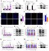
Figure 1
Figure 1
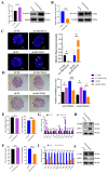
SLCO3A1 promotes the proliferation of GCs. (A) RT-qPCR analysis of SLCO3A1 expression in GCs transfected with different concentrations of pcDNA3.1 (OE-NC) or pcDNA3.1-SLCO3A1 (OE-SLCO3A1). (B) Western blot analysis of SLCO3A1 transfected with 0.5 ng/µL of OE-NC or OE-SLCO3A1. (C) RT-qPCR analysis of SLCO3A1 expression in GCs transfected with 50 nM or 100 nM of siRNAs (siRNA#NC, siRNA#1, siRNA#2, siRNA#3). (D) Western blot analysis of SLCO3A1 transfected with 50 nM of siRNA#3. EdU detection of proliferation rate transfected with OE-SLCO3A1 (E) or KD-SLCO3A1 (F). Scale bar = 200 μm. RT-qPCR analysis of proliferation-related genes transfected with OE-SLCO3A1 (G) or KD-SLCO3A1 (I). Western blot analysis of MCL-1, PCNA and STAR transfected with OE-SLCO3A1 (H) or KD-SLCO3A1 (J). RT-qPCR analysis of estrogen pathway-related genes transfected with OE-SLCO3A1 (K) and KD-SLCO3A1 (M). Western blot analysis of FSHR transfected with OE-SLCO3A1 (L) and KD-SLCO3A1 (N). All of the above experimental results were obtained in the COV434 cells. * p < 0.05, ** p < 0.01, and *** p < 0.001.
Figure 2
Figure 2
SLCO3A1 promotes the cell cycle-related processes but inhibits the apoptosis of GCs. The cell cycle distribution was analyzed by flow cytometric transfected with OE-SLCO3A1 (A) or KD-SLCO3A1 (B). Green: cells in the G0-G1 phase, Yellow: cells in S phase, Blue: cells in G2-M phase. (C) RT-qPCR analysis of cycle-related genes expression transfected with OE-NC or OE-SLCO3A1. (D) Western blot analysis of CCNE1, CDK2 and CCND1 transfected with OE-NC or OE-SLCO3A1. (E) RT-qPCR analysis of cycle-related genes expression transfected with KD-NC or KD-SLCO3A1. (F) Western blot analysis of CCNE1, CDK2 and CCND1 transfected with KD-NC or KD-SLCO3A1. Flow cytometry assay of the apoptosis rate transfected with OE-SLCO3A1 (G) or KD-SLCO3A1 (H). Q1: dead cells and cell debris, Q2: late apoptotic cells, Q3: early apoptotic cells, Q4: negative cells. RT-qPCR analysis of pro-apoptosis related genes expression transfected with OE-SLCO3A1 (I) or KD-SLCO3A1 (K). Western blot analysis of CASP3 and CASP8 transfected with OE-SLCO3A1 (J) or KD-SLCO3A1 (L). All of the above experimental results were obtained in the COV434 cells. * p < 0.05, ** p < 0.01, *** p < 0.001, ns p > 0.05.
Figure 3
Figure 3
SLCO3A1 promotes the growth of porcine follicles. The mRNA and protein levels of SLCO3A1 of porcine follicles treated with LV-SLCO3A1 (A) or KD-SLOC3A1 (B). RT-qPCR analysis of proliferation- and cycle-related genes expression treated with LV-SLCO3A1 (C) or sh-SLCO3A1 (E). Western blot analysis of P65 and CDK4 treated with LV-SLCO3A1 (D) or sh-SLCO3A1 (F). * p < 0.05, ** p < 0.01, *** p < 0.001, ns p > 0.05.
Figure 4
Figure 4
SLCO3A1 promotes follicular growth in mice. The mRNA and protein levels of SLCO3A1 in the ovaries of mice treated with LV-SLCO3A1 (A) and sh-SLCO3A1 (B). (C) The TUNEL staining was performed in mice ovaries treated with LV-SLCO3A1 or sh-SLCO3A1, and the relative fluorescence intensity was corrected by that of the control groups. Yellow arrows indicated apoptotic GCs. Scale bar = 500 μm. (D) The HE staining and follicular statistics of mice after their ovaries were treated with LV-SLCO3A1 or sh-SLCO3A1. Black arrows indicated antral follicles, and orange arrows indicated corpus luteum. Scale bar = 500 μm. (E) The age of mice at vaginal opening treated with LV-SLCO3A1 or sh-SLCO3A1. (F) The concentration of E2 treated with LV-SLCO3A1 or sh-SLCO3A1 was assessed by ELISA. RT-qPCR analysis of proliferation- and cycle-related genes expression treated with LV-SLCO3A1 (G) or sh-SLCO3A1 (I). Western blot analysis of CCNB2 and CDK1 treated with LV-SLCO3A1 (H) or sh-SLCO3A1 (J). * p < 0.05, ** p < 0.01, *** p < 0.001.
Figure 5
Figure 5
Knockdown of DNMT1 upregulates the mRNA and protein levels of SLCO3A1. (A) RT-qPCR analysis of SLCO3A1 expression in the small (<3 mm in diameter) and large (>3 mm in diameter) follicles. (B) Western blot analysis of SLCO3A1 in the small and large follicles. (C) RT-qPCR analysis of SLCO3A1 expression in the DMSO or 5-Aza-CdR treated-cells. (D) Western blot analysis of SLCO3A1 in the DMSO or 5-Aza-CdR treated-cells. (E) Chromatin accessibility assay of region-1, region-2, and region-3 in the SLCO3A1 promoter treated with DMSO or 5-Aza-CdR (region-1, +971/+1252 bp, region-2, +1433/+1557 bp, and region-3, +1623/+1831 bp, transcription start site = +2000 bp). (F) RT-qPCR analysis of SLCO3A1 expression in GCs transfected with siRNAs of DNMT1, DNMT3A, or DNMT3B. Western blot analysis of SLCO3A1, DNMT1 (G), DNMT3A (H), and DNMT3B (I) in the siRNAs of DNMT1, DNMT3A, or DNMT3B treated-cells. *** p < 0.001.

Similar articles

Cited by

References

    1. Murase T., Iwase A., Komatsu K., Bayasula, Nakamura T., Osuka S., Takikawa S., Goto M., Kotani T., Kikkawa F. Follicle Dynamics: Visualization and Analysis of Follicle Growth and Maturation Using Murine Ovarian Tissue Culture. J. Assist. Reprod. Genet. 2018;35:339–343. doi: 10.1007/s10815-017-1073-5. - DOI - PMC - PubMed
    1. Li C., Liu Z., Wu G., Zang Z., Zhang J.-Q., Li X., Tao J., Shen M., Liu H. FOXO1 Mediates Hypoxia-Induced G0/G1 Arrest in Ovarian Somatic Granulosa Cells by Activating the TP53INP1-P53-CDKN1A Pathway. Development. 2021;148:dev199453. doi: 10.1242/dev.199453. - DOI - PubMed
    1. Zeng X., Xie Y., Liu Y., Long S., Mo Z. Polycystic Ovarian Syndrome: Correlation between Hyperandrogenism, Insulin Resistance and Obesity. Clin. Chim. Acta. 2020;502:214–221. doi: 10.1016/j.cca.2019.11.003. - DOI - PubMed
    1. Park E., Cockrem J.F., Han K.-H., Kim D.-H., Jung M.-H., Chu J.-P. Stress-Induced Activation of Ovarian Heat Shock Protein 90 in a Rat Model of Polycystic Ovary Syndrome. J. Obstet. Gynaecol. Res. 2012;38:396–407. doi: 10.1111/j.1447-0756.2011.01705.x. - DOI - PubMed
    1. Fan W., Yuan Z., Li M., Zhang Y., Nan F. Decreased Oocyte Quality in Patients with Endometriosis Is Closely Related to Abnormal Granulosa Cells. Front. Endocrinol. 2023;14:1226687. doi: 10.3389/fendo.2023.1226687. - DOI - PMC - PubMed

LinkOut - more resources