. 2024 Feb 21;25(5):2503.
doi: 10.3390/ijms25052503.

Enhanced Diaphragm Muscle Function upon Satellite Cell Transplantation in Dystrophic Mice

Affiliations

Enhanced Diaphragm Muscle Function upon Satellite Cell Transplantation in Dystrophic Mice

Karim Azzag et al. Int J Mol Sci. .

Abstract

The diaphragm muscle is essential for breathing, and its dysfunctions can be fatal. Many disorders affect the diaphragm, including muscular dystrophies. Despite the clinical relevance of targeting the diaphragm, there have been few studies evaluating diaphragm function following a given experimental treatment, with most of these involving anti-inflammatory drugs or gene therapy. Cell-based therapeutic approaches have shown success promoting muscle regeneration in several mouse models of muscular dystrophy, but these have focused mainly on limb muscles. Here we show that transplantation of as few as 5000 satellite cells directly into the diaphragm results in consistent and robust myofiber engraftment in dystrophin- and fukutin-related protein-mutant dystrophic mice. Transplanted cells also seed the stem cell reservoir, as shown by the presence of donor-derived satellite cells. Force measurements showed enhanced diaphragm strength in engrafted muscles. These findings demonstrate the feasibility of cell transplantation to target the diseased diaphragm and improve its contractility.

Keywords: diaphragm; engraftment; muscle stem cells; muscular dystrophy; pre-injury; regeneration; satellite cells; specific force; transplantation.


Conflict of interest statement

R.C.R.P. is a cofounder of and holds equity in Myogenica. All other authors have no competing financial interests.

Figures
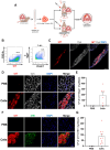
Figure 1
Figure 1
Detection of donor-derived myofibers in diaphragm muscles of NSG and NSG-FKRPP448L mice. (A) Schematic representation outlining the isolation of satellite cells (SCs) from the diaphragm of a mTmG mouse and their transplantation into the diaphragm muscles of NSG and NSG-FKRPP448L mice. (B) Representative FACS plots show SC isolation from mTmG mice based on the expression of Itga7 and Vcam1, following gating of the tdT+Lin- fraction. (CE) Diaphragm transplantation in NSG mice. Representative images show the whole area of engraftment, as indicated by immunostaining for tdT (in red), dystrophin (Dys) (in gray), and DAPI (in blue). Scale bar is 200 µm (C). Representative images show the same staining at higher magnification for PBS- (upper panel) and SC-injected (lower panel) diaphragm muscles. Scale bar is 50 µm (D). Graph shows quantification of engraftment ((E) from (D)) based on the number of tdT+Dys+ myofibers. Data are shown as mean ± SEM (n = 3 for PBS and n = 6 for cells). (F,G) Diaphragm transplantation in NSG-FKRPP448L mice. Representative images show staining for tdT and IIH6 in PBS- (upper panel) and SC-injected (lower panel) diaphragm muscles. Scale bar is 50 µm (F). Respective quantification of engraftment (G), as shown by the number of tdT+IIH6+ myofibers. Data are shown as mean ± SEM (n = 3 for PBS and n = 10 for cells).
Figure 2
Figure 2
Engraftment of SCs isolated from Pax7-ZsGreen–mTmG mice in the diaphragm of NSG-FKRPP448L mice. (A) Scheme outlines the isolation of SCs from Pax7-ZsGreen–mTmG mice and their transplantation into diaphragm muscles of NSG-FKRPP448L mice. (B) Representative FACS plots show SC isolation from Pax7-ZsGreen–mTmG mice based on the expression of ZsGreen. (C,D) Representative images show diaphragm engraftment as indicated by immunostaining for tdT (in red), IIH6 (in green), and DAPI (in blue). Scale bar is 200 µm (C). (D) shows higher magnification of control (upper panel) and transplanted (lower panel) diaphragms. Scale bar is 50 µm. (E) Graph shows engraftment quantification (D), as shown by the number of tdT+IIH6+ myofibers. Data are shown as mean ± SEM (n = 4 for PBS and n = 9 for cells).
Figure 3
Figure 3
Rescue of dystrophin expression upon SC transplantation into the diaphragm of NSG-mdx4Cv mice. (A) Schematic representation of transplantation outline. (B) Representative images show the presence of tdT+Dys+ (red and gray, respectively) myofibers in only the transplanted diaphragm muscles (lower panel). DAPI in blue-stained nuclei. Scale bar is 50 µm. (C) Graph shows respective engraftment quantification (B). Data are shown as mean ± SEM (n = 2 for PBS and n = 4 for cells).
Figure 4
Figure 4
Diaphragm engraftment results in improvement of muscle force in mdx4Cv mice. (A) Scheme indicates the outline of transplantation into the diaphragm muscles of immunocompetent mdx4Cv mice undergoing tacrolimus immunosuppression. One month later, the diaphragm was assessed for engraftment and force measurement. (B) Representative images show immunostaining for tdT (in red), Dys (in gray), and DAPI (blue) for PBS-injected (top panel) and cell-injected (lower panel) diaphragms. Scale bar is 50 µm. (C) Specific (sF0: maximum force normalized to CSA) force of cell-injected diaphragm compared with PBS. Data are shown as mean ± SEM (n = 7 for PBS and cells). * p < 0.05 according to t test. (D) Specific force measured at different stimulation frequencies, comparing cells with PBS. Data are shown as mean ± SEM (n = 7 for each group). * p < 0.05 according to t test. (E) Representative images show engraftment in a cell-injected diaphragm strip, as indicated by the presence of tdT (in red). Brightfield is indicated in gray. Scale bar is 100 µm. (F) Graph shows quantification of the number of tdT+ myofibers present in transplanted diaphragm strips. Data are shown as mean ± SEM (n = 7 for each group).

References

    1. Ricoy J., Rodríguez-Núñez N., Álvarez-Dobaño J.M., Toubes M.E., Riveiro V., Valdés L. Diaphragmatic dysfunction. Pulmonology. 2019;25:223–235. doi: 10.1016/j.pulmoe.2018.10.008. - DOI - PubMed
    1. Mauro A.L., Aliverti A. Physiology of respiratory disturbances in muscular dystrophies. Breathe. 2016;12:318–327. doi: 10.1183/20734735.012716. - DOI - PMC - PubMed
    1. Richard I., Hogrel J., Stockholm D., Payan C.A.M., Fougerousse F., Eymard B., Mignard C., de Munain A.L., Fardeau M., Urtizberea J.A., et al. Natural history of LGMD2A for delineating outcome measures in clinical trials. Ann. Clin. Transl. Neurol. 2016;3:248–265. doi: 10.1002/acn3.287. - DOI - PMC - PubMed
    1. Wohlgemuth M., van der Kooi E.L., van Kesteren R.G., van der Maarel S.M., Padberg G.W. Ventilatory support in facioscapulohumeral muscular dystrophy. Neurology. 2004;63:176–178. doi: 10.1212/01.WNL.0000133126.86377.E8. - DOI - PubMed
    1. Mathieu J., Allard P., Potvin L., Prévost C., Bégin P. A 10-year study of mortality in a cohort of patients with myotonic dystrophy. Neurology. 1999;52:1658–1662. doi: 10.1212/WNL.52.8.1658. - DOI - PubMed

LinkOut - more resources