. 2024 Feb 21;25(5):2505.
doi: 10.3390/ijms25052505.

Fibroblast Growth Factor Receptor Inhibitors Decrease Proliferation of Melanoma Cell Lines and Their Activity Is Modulated by Vitamin D

Affiliations

Fibroblast Growth Factor Receptor Inhibitors Decrease Proliferation of Melanoma Cell Lines and Their Activity Is Modulated by Vitamin D

Anna Piotrowska et al. Int J Mol Sci. .

Abstract

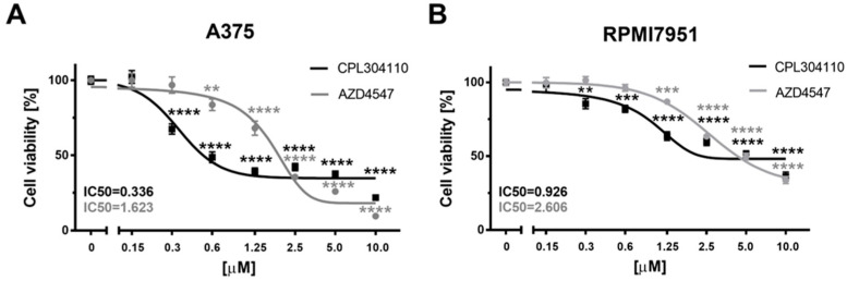
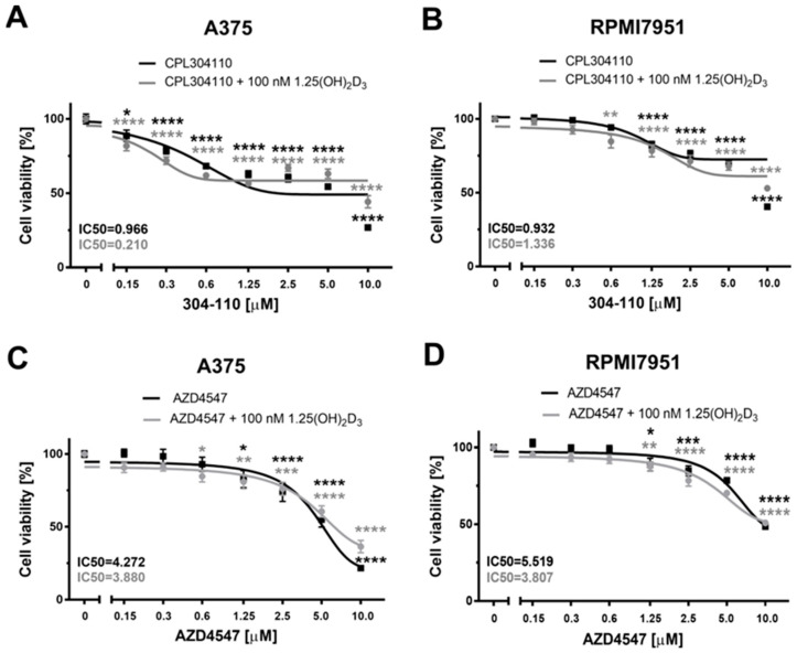
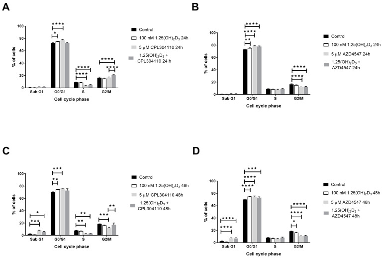
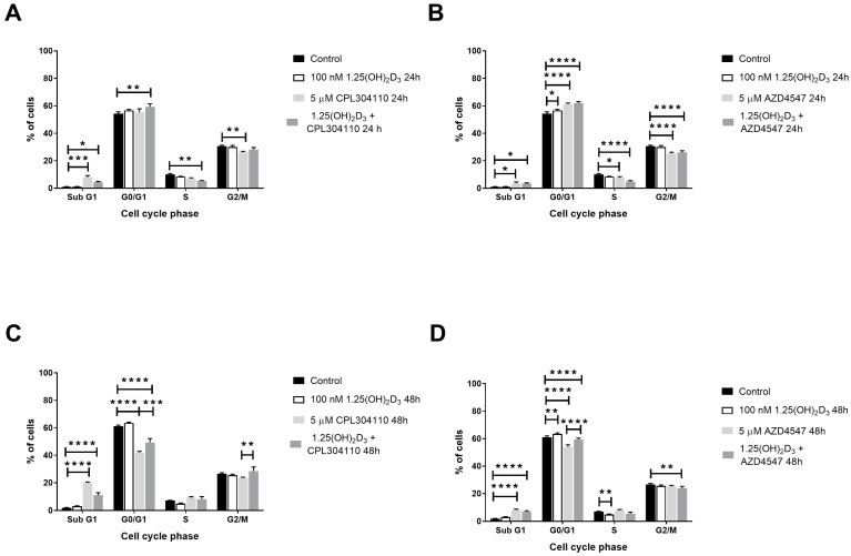
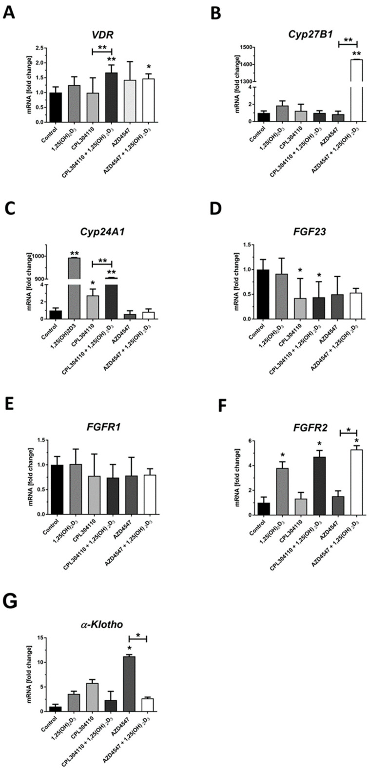
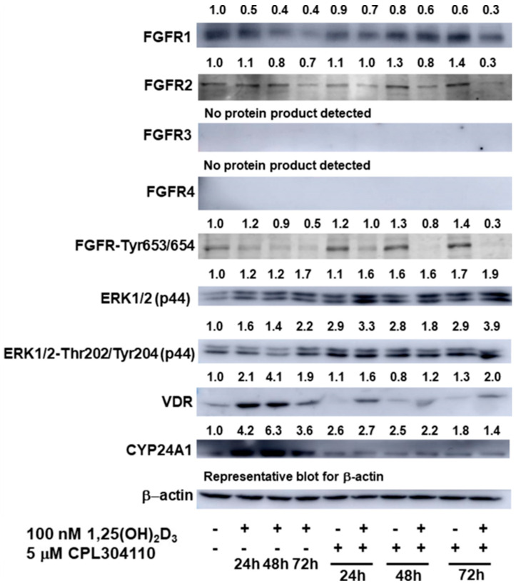
Regardless of the unprecedented progress in malignant melanoma treatment strategies and clinical outcomes of patients during the last twelve years, this skin cancer remains the most lethal one. We have previously documented that vitamin D and its low-calcaemic analogues enhance the anticancer activity of drugs including a classic chemotherapeutic-dacarbazine-and an antiangiogenic VEGFRs inhibitor-cediranib. In this study, we explored the response of A375 and RPMI7951 melanoma lines to CPL304110 (CPL110), a novel selective inhibitor of fibroblast growth factor receptors (FGFRs), and compared its efficacy with that of AZD4547, the first-generation FGFRs selective inhibitor. We also tested whether 1,25(OH)2D3, the active form of vitamin D, modulates the response of the cells to these drugs. CPL304110 efficiently decreased the viability of melanoma cells in both A375 and RPMI7951 cell lines, with the IC50 value below 1 µM. However, the metastatic RPMI7951 melanoma cells were less sensitive to the tested drug than A375 cells, isolated from primary tumour site. Both tested FGFR inhibitors triggered G0/G1 cell cycle arrest in A375 melanoma cells and increased apoptotic/necrotic SubG1 fraction in RPMI7951 melanoma cells. 1,25(OH)2D3 modulated the efficacy of CPL304110, by decreasing the IC50 value by more than 4-fold in A375 cell line, but not in RPMI7951 cells. Further analysis revealed that both inhibitors impact vitamin D signalling to some extent, and this effect is cell line-specific. On the other hand, 1,25(OH)2D3, have an impact on the expression of FGFR receptors and phosphorylation (FGFR-Tyr653/654). Interestingly, 1,25(OH)2D3 and CPL304110 co-treatment resulted in activation of the ERK1/2 pathway in A375 cells. Our results strongly suggested possible crosstalk between vitamin D-activated pathways and activity of FGFR inhibitors, which should be considered in further clinical studies.

Keywords: 1,25(OH)2D; AZD4547; CPL304110; FGFR; FGFR inhibitors; melanoma; vitamin D.


Conflict of interest statement

Authors D.P. and M.W. was employed by the company Celon Pharma S.A. The remaining authors declare that the research was conducted in the absence of any commercial or financial relationships that could be construed as a potential conflict of interest.

Figures

Figure 1
Figure 1
The effect of FGFR inhibitors, CPL304110 and AZD4547, on the viability of human malignant A375 (A) and RPMI7951 (B) melanoma cell lines. A375 melanoma cells were treated with serial dilutions (0.15–10 µM) of CPL304110 or AZD4547 for 72 h. Data are shown as the mean of three independent experiments (n = 4–6 in each) ± SEM. Statistical significance for a single treatment was estimated using one-way ANOVA and presented as ** p < 0.01, *** p < 0.001 or **** p < 0.0001.
Figure 2
Figure 2
The effect of FGFR inhibitors, CPL304110 (A,B) and AZD4547 (C,D), and their combinations with 1,25(OH)2D3 on viability of the human malignant A375 (A,C) and RPMI7951 (B,D) melanoma cell lines. Cells were treated with serial dilutions (0.15–10 µM) of CPL304110 or AZD4547 alone or in combination with 1,25(OH)2D3 at 100 nM concentration for 48 h. Data are shown as the mean of three independent experiments (n = 4–6 in each) ± SEM. Statistical significance for single treatment was estimated using one-way ANOVA and presented as * p < 0.05, ** p < 0.01, *** p < 0.001 or **** p < 0.0001.
Figure 3
Figure 3
The effect of FGFR inhibitors, CPL304110 (A,C) and AZD4547 (B,D) at 5 µM or its combination with 100 nM 1,25(OH)2D3 on the distribution of human malignant melanoma A375 throughout phases of the cell cycle (SubG1—apoptotic/necrotic cells, G1—growth, S—DNA synthesis, G2/M—preparation for mitosis/mitosis) after 24 or 48 h incubation. Cells were harvested, stained with propidium iodide and analysed by Flow Cytometry. The same control and 1,25(OH)2D3 data are plotted in an appropriate incubation time graph (for 24 h—(A,B); and for 48 h—(C,D)). The data are presented as the mean ± standard deviation (n = 3). Statistical significance was estimated using two-way ANOVA followed by Tukey’s multiple comparison test and presented as * p < 0.05; ** p < 0.01; *** p < 0.001, **** p < 0.0001.
Figure 4
Figure 4
The effect of FGFR inhibitors, CPL304110 (A,C) and AZD4547 (B,D) at 5 µM or their combination with 100 nM 1,25(OH)2D3 on the distribution of human malignant melanoma RPMI7951, throughout phases of the cell cycle (SubG1—apoptotic/necrotic cells, G1—growth, S—DNA synthesis, G2/M—preparation for mitosis/mitosis) after 24 or 48 h incubation. Cells were harvested, stained with propidium iodide and analysed by Flow Cytometry. The same control and 1,25(OH)2D3 data are plotted on an appropriate incubation time graph (for 24 h—(A,B); and for 48 h—(C,D)). The data are presented as the mean ± standard deviation (n = 3). Statistical significance was estimated using two-way ANOVA followed by Tukey’s multiple comparison test and presented as * p < 0.05; ** p < 0.01; *** p < 0.001, **** p < 0.0001.
Figure 5
Figure 5
The effect of FGFR inhibitors, CPL304110 and AZD4547, at 5 µM, or their combination with 100 nM 1,25(OH)2D3 on VDR (A), CYP27B1 (B), CYP24A1 (C), FGF23 (D), FGFR1 (E), FGFR2 (F) and α-KLOTHO (KL) (G) gene expression in A375 after 24 h incubation. mRNA levels were measured by qPCR. The results are representative of three experiments carried out in duplicate. * p < 0.05, ** p < 0.01 and *** p < 0.001 vs. untreated control or between the two groups indicated by the bracket.
Figure 6
Figure 6
The effect of FGFR inhibitors, CPL304110 and AZD4547, at 5 µM, or their combination with 100 nM 1,25(OH)2D3 on VDR (A), CYP27B1 (B), CYP24A1 (C), FGF23 (D), FGFR1 (E), FGFR2 (F) and α-KLOTHO (KL) (G) gene expression in RPMI7951 after 24 h incubation. mRNA levels were measured by qPCR. The results are representative of three experiments carried out in duplicate. * p < 0.05 and ** p < 0.01 vs. untreated control or between the two groups indicated by the bracket.
Figure 7
Figure 7
The effect of FGFR inhibitor CPL304110 at 5 µM, or its combination with 1,25(OH)2D3 at 100 nM concentration on FGFR1-4, phosphorylated at Tyr653/654 FGFR, ERK1/2 (p44), phosphorylated at Thr202/Tyr202 ERK1/2 (p44), VDR and CYP24A1 protein level in A375 during 24 h–72 h incubation. Protein levels were measured by Western blotting, with β-actin used as a control. Data are shown as representative blots from three independent experiments. The visualised bands’ densitometric values presented above are calculated as ratios to appropriate β-actin (not shown in Figure) and are further normalised to nontreated “control” samples, which are referred as “1,0”. β-actin blot presented in the figure is representative.

Similar articles

Cited by

References

    1. Garbe C., Amaral T., Peris K., Hauschild A., Arenberger P., Basset-Seguin N., Bastholt L., Bataille V., Del Marmol V., Dréno B., et al. European consensus-based interdisciplinary guideline for melanoma. Part 1: Diagnostics: Update 2022. Eur. J. Cancer. 2022;170:236–255. doi: 10.1016/j.ejca.2022.03.008. - DOI - PubMed
    1. Siegel R.L., Giaquinto A.N., Jemal A. Cancer statistics, 2024. CA Cancer J. Clin. 2024;74:12–49. doi: 10.3322/caac.21820. - DOI - PubMed
    1. Siegel R.L., Miller K.D., Fuchs H.E., Jemal A. Cancer Statistics, 2021. CA Cancer J. Clin. 2021;71:7–33. doi: 10.3322/caac.21654. - DOI - PubMed
    1. Siegel R.L., Miller K.D., Fuchs H.E., Jemal A. Cancer statistics, 2022. CA Cancer J. Clin. 2022;72:7–33. doi: 10.3322/caac.21708. - DOI - PubMed
    1. Ascierto P.A., Puzanov I., Agarwala S.S., Blank C., Carvajal R.D., Demaria S., Dummer R., Ernstoff M., Ferrone S., Fox B.A., et al. Perspectives in melanoma: Meeting report from the “Melanoma Bridge” (December 5th–7th, 2019, Naples, Italy) J. Transl. Med. 2020;18:346. doi: 10.1186/s12967-020-02482-x. - DOI - PMC - PubMed