Review
. 2024 Feb 21;25(5):2530.
doi: 10.3390/ijms25052530.

Membrane-Driven Dimerization of the Peripheral Membrane Protein KRAS: Implications for Downstream Signaling

Affiliations
Review

Membrane-Driven Dimerization of the Peripheral Membrane Protein KRAS: Implications for Downstream Signaling

Ki-Young Lee. Int J Mol Sci. .

Abstract

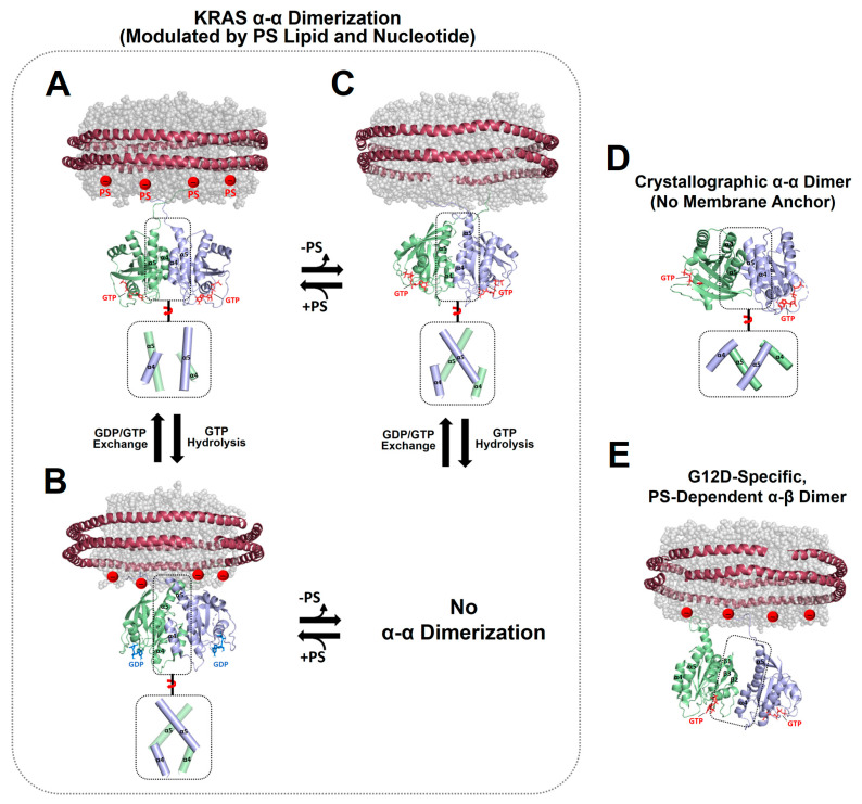
Transient homo-dimerization of the RAS GTPase at the plasma membrane has been shown to promote the mitogen-activated protein kinase (MAPK) signaling pathway essential for cell proliferation and oncogenesis. To date, numerous crystallographic studies have focused on the well-defined GTPase domains of RAS isoforms, which lack the disordered C-terminal membrane anchor, thus providing limited structural insight into membrane-bound RAS molecules. Recently, lipid-bilayer nanodisc platforms and paramagnetic relaxation enhancement (PRE) analyses have revealed several distinct structures of the membrane-anchored homodimers of KRAS, an isoform that is most frequently mutated in human cancers. The KRAS dimerization interface is highly plastic and altered by biologically relevant conditions, including oncogenic mutations, the nucleotide states of the protein, and the lipid composition. Notably, PRE-derived structures of KRAS homodimers on the membrane substantially differ in terms of the relative orientation of the protomers at an "α-α" dimer interface comprising two α4-α5 regions. This interface plasticity along with the altered orientations of KRAS on the membrane impact the accessibility of KRAS to downstream effectors and regulatory proteins. Further, nanodisc platforms used to drive KRAS dimerization can be used to screen potential anticancer drugs that target membrane-bound RAS dimers and probe their structural mechanism of action.

Keywords: KRAS; dimerization; nanodisc; paramagnetic relaxation enhancement (PRE); peripheral membrane protein.


Conflict of interest statement

The author declares no conflicts of interest.

Figures

Figure 2
Figure 2
Representative NMR-driven structures of symmetric α–α dimers of full-length KRAS in the GTP-bound (A) and GDP-bound (B) states on anionic membranes containing phosphatidylserine (PS) lipids (PDB IDs: 6W4E and 6W4F) and the α–α dimer of GTP-bound KRAS on a neutral membrane lacking PS (C). The dimerization of GDP-bound KRAS is unfavorable on the neutral membrane. (D) Crystal structure of a symmetric α–α dimer of membrane-free KRAS lacking the disordered C-terminal hypervariable region (HVR) responsible for membrane association (PDB ID: 5VQ2). (E) The representative NMR-driven structure of an asymmetric α–β dimer of the full-length KRAS-G12D mutant on an anionic membrane containing PS (PDB ID: 7RSE). This mutant also forms the symmetric α–α dimer shared with wild-type KRAS. Differences in the relative orientation of the protomers at the α–α dimer interfaces are highlighted in dotted boxes.
Figure 3
Figure 3
Plasticity of the KRAS dimer interface. (A) Overlay between representative NMR-driven structures of full-length KRAS-GTP dimers on both anionic (phosphatidylserine [PS]-enriched) and neutral (PS-free) membranes and the full-length KRAS-GDP dimer on the anionic membrane (PDB IDs: 6W4E and 6W4F). For clarity, only the GTPase domains (residues 1–172) of the structures are shown. One protomer in each KRAS dimer is overlaid to clearly show the different arrangements of the opposing protomers. (B) Overlay between the crystal structures of membrane-free KRAS dimers in both GTP- and GDP-bound states lacking the disordered C-terminal hypervariable region (HVR) (PDB IDs: 5VQ2 and 5W22). (C) Promiscuous intermolecular interactions at the α–α dimer interface of full-length KRAS on the membrane. The KRAS dimer interfaces involve intermolecular electrostatic interactions that are modulated by the nucleotide-bound state of KRAS and the lipid composition of the membrane. Representative electrostatic interactions with a cut-off distance < 3.5 Å at the dimer interface, validated by interface-specific mutagenesis, are schematically indicated.
Figure 4
Figure 4
Overlay of representative NMR-derived structures of KRAS-GTP dimers on membranes containing or lacking phosphatidylserine (PS) with the structures of KRAS in complex with the autoinhibited BRAF–14-3-3 complex (A) and the RAS binding domain (RBD)–cysteine rich domain (CRD) of active BRAF (B). The arrow represents steric clash or charge repulsion between the effector and the membrane surface. The complex models are reconstituted based on the cryo-electron microscopy (EM) structure of the BRAF–14-3-3 complex (PDB ID: 7MFE), the crystal structure of KRAS in complex with the RBD–CRD (PDB ID: 6XI7), and the NMR-derived structure of the KRAS-GTP dimer on the membrane (PDB ID: 6W4E).
Figure 5
Figure 5
Overlay of representative NMR-derived structures of KRAS-GTP dimers on membranes containing or lacking phosphatidylserine (PS) with the structures of KRAS in complex with phosphoinositide 3-kinase gamma (PI3Kγ). The arrow represents steric clash between the effector and the membrane surface. The complex models are reconstituted based on the crystal structures of KRAS in complex with PI3Kγ (PDB ID: 1HE8) and the NMR-derived structure of the KRAS-GTP dimer on the membrane (PDB ID: 6W4E).
Figure 6
Figure 6
Structural models of membrane-bound KRAS dimers in complex with the catalytic domains of son of sevenless homolog 1 (SOS1) and Ras GTPase activating protein (RasGAP). Representative NMR-derived structures of KRAS-GTP dimers on membranes containing or lacking phosphatidylserine (PS) are overlaid with the crystal structure of KRAS in complex with the catalytic domain of RasGAP (PDB ID: 1WQ1). The representative NMR-derived structure of the KRAS-GDP dimer on an anionic membrane containing PS is overlaid with the crystal structure of KRAS in complex with the REM–CDC25 domain of SOS1 (PDB ID: 6EPL).
Figure 7
Figure 7
The structural mechanism of KRAS dimerization modulators. (A) Overlay of representative NMR-driven structures of KRAS α–α dimers with the crystal structures of KRAS in complex with the following synthetic proteins: the NS1 monobody (PDB ID: 5E95) and designed ankyrin repeat proteins (DARPins) K13 (PDB ID: 6H46) and K19 (PDB ID: 6H47). These KRAS binders cause steric clashes with KRAS protomers in dimers, as indicated by the arrows, and act as competitive inhibitors of KRAS dimerization. One protomer in each KRAS dimer is overlaid to clearly show the different arrangements of the opposing protomers. (B) BI2852-mediated formation of an inactive β-β dimer of KRAS-G12D (PDB ID: 6GJ8) in which the effector binding sites at the β-interfaces are inaccessible to RAS effectors. (C) Overlay of the representative NMR-driven structure of membrane-anchored KRAS in complex with the protein–membrane interaction (PMI) stabilizer Cmpd2 (PDB ID: 6CC9) with representative NMR-driven structures of KRAS-GTP α–α dimers on phosphatidylserine (PS)-enriched anionic or PS-free neutral membranes and the KRAS-GDP α–α dimer on a PS-enriched anionic membrane. Red arrows represent the positions to which the disordered C-terminal membrane anchors (K172-C185) of KRAS are attached.
Figure 1
Figure 1
The structure and functional sites of KRAS. (A) Crystal structure of KRAS in the active GTPγS (a non-hydrolyzable GTP analog)-bound state (PDB ID: 4DSO). (B) Schematic of functional sites of KRAS. KRAS dimerization involves several different ‘α4–α5’ surfaces, as determined using NMR and X-ray crystallography, and the β-sheet region including Switch I and II sites binds to RAS effectors (e.g., RAF, PI3Kγ) or regulators (e.g., GAP, GEF). (C) The amino acid sequence of KRAS. Residues 12, 13, and 61, whose single-point mutations are associated with many human cancers, are indicated as black arrows. Secondary structure content is indicated at the bottom of the sequence with α-helices (blue) and β-strands (yellow). Nucleotide binds to a conserved phosphate-binding loop (P-loop comprising residues 10–17, green). Switch I (residues 30–40) and Switch II (residues 58–72) regions and disordered C-terminal polybasic region (PBR, residues 172–184) are colored red, violet, and blue, respectively.

Similar articles

Cited by

References

    1. Tan S., Tan H.T., Chung M.C. Membrane proteins and membrane proteomics. Proteomics. 2008;8:3924–3932. doi: 10.1002/pmic.200800597. - DOI - PubMed
    1. Yin H., Flynn A.D. Drugging Membrane Protein Interactions. Annu. Rev. Biomed. Eng. 2016;18:51–76. doi: 10.1146/annurev-bioeng-092115-025322. - DOI - PMC - PubMed
    1. Ahram M., Litou Z.I., Fang R., Al-Tawallbeh G. Estimation of membrane proteins in the human proteome. Silico Biol. 2006;6:379–386. - PubMed
    1. Cournia Z., Allen T.W., Andricioaei I., Antonny B., Baum D., Brannigan G., Buchete N.V., Deckman J.T., Delemotte L., del Val C., et al. Membrane Protein Structure, Function, and Dynamics: A Perspective from Experiments and Theory. J. Membr. Biol. 2015;248:611–640. doi: 10.1007/s00232-015-9802-0. - DOI - PMC - PubMed
    1. Saad S., Jarosz D.F. Protein self-assembly: A new frontier in cell signaling. Curr. Opin. Cell Biol. 2021;69:62–69. doi: 10.1016/j.ceb.2020.12.013. - DOI - PMC - PubMed

LinkOut - more resources