. 2024 Feb 22;25(5):2545.
doi: 10.3390/ijms25052545.

Dihydropashanone Isolated from Lindera erythrocarpa, a Potential Natural Product for the Treatment of Neurodegenerative Diseases

Affiliations

Dihydropashanone Isolated from Lindera erythrocarpa, a Potential Natural Product for the Treatment of Neurodegenerative Diseases

Zhiming Liu et al. Int J Mol Sci. .

Abstract

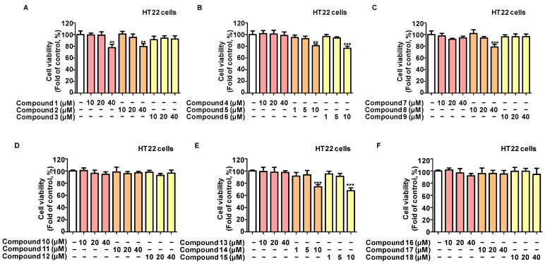
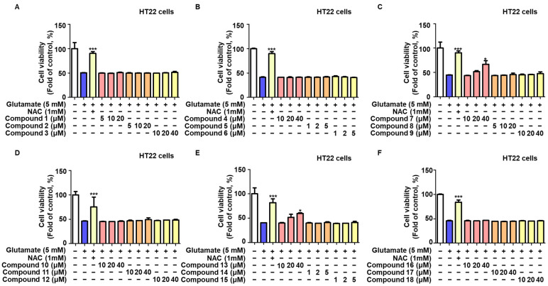
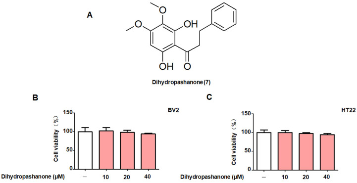
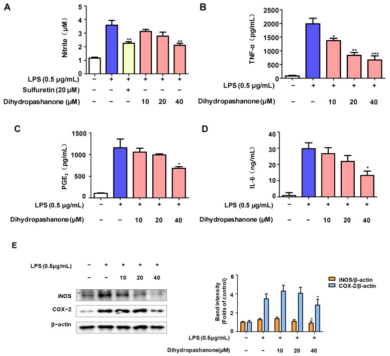
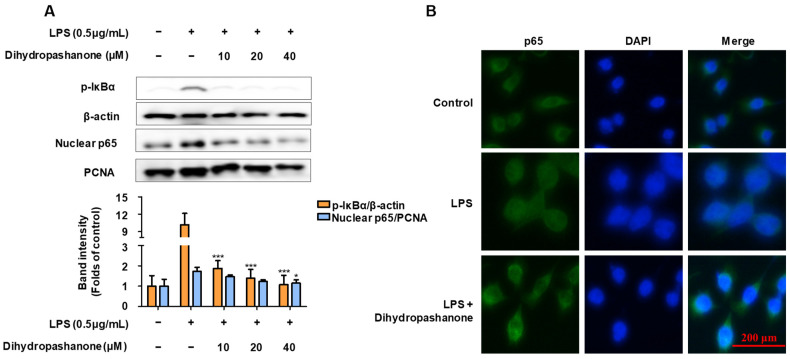
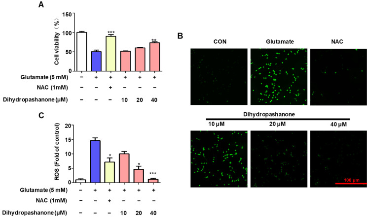
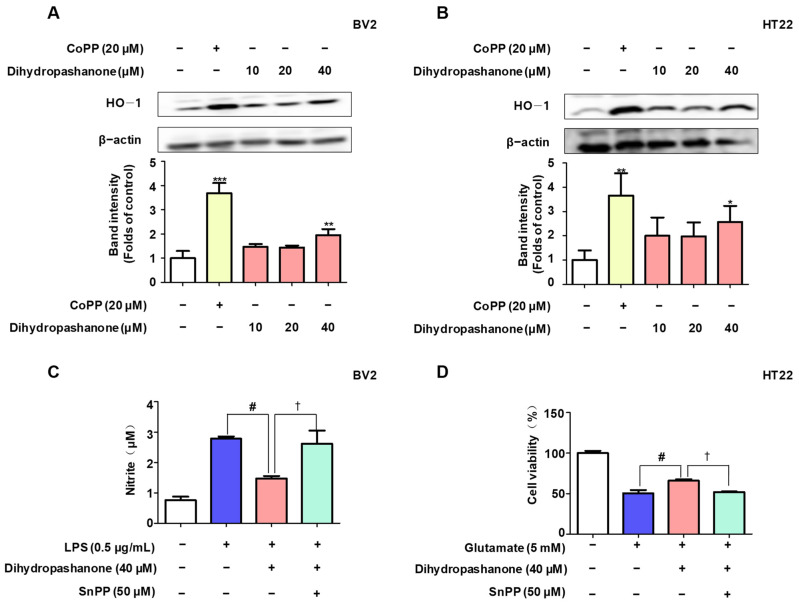
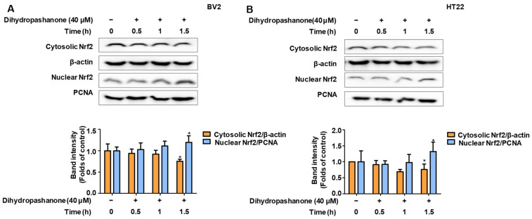
Lindera erythrocarpa, a flowering plant native to eastern Asia, has been reported to have neuroprotective activity. However, reports on the specific bioactive compounds in L. erythrocarpa are finite. The aim of this study was to investigate the anti-neuroinflammatory and neuroprotective effects of the compounds isolated from L. erythrocarpa. Dihydropashanone, a compound isolated from L. erythrocarpa extract, was found to have protected mouse hippocampus HT22 cells from glutamate-induced cell death. The antioxidant and anti-inflammatory properties of dihydropashanone in mouse microglial BV2 and HT22 cells were explored in this study. The results reveal that dihydropashanone inhibits lipopolysaccharide-induced inflammatory response and suppresses the activation of nuclear factor (NF)-κB in BV2 cells. In addition, dihydropashanone reduced the buildup of reactive oxygen species in HT22 cells and induced activation of the nuclear factor E2-related factor 2 (Nrf2)/heme oxygenase (HO)-1 signaling pathway in BV2 and HT22 cells. Our results suggest that dihydropashanone reduces neuroinflammation by decreasing NF-κB activation in microglia cells and protects neurons from oxidative stress via the activation of the Nrf2/HO-1 pathway. Thus, our data suggest that dihydropashanone offers a broad range of applications in the treatment of neurodegenerative illnesses.

Keywords: NF-κB; Nrf2; dihydropashanone; neuroprotective effects.


Conflict of interest statement

The authors declare no conflicts of interest.

Figures

Figure 1
Figure 1
Cytotoxicity of the 18 compounds on HT22 cells. HT22 cells were treated with varying doses of the compounds for 24 h, and cell viability (AF) was assessed using the MTT test. Values are reported as % of control and presented as the mean ± standard deviation (SD) of at least three independent experiments. ** p < 0.01, *** p < 0.001 vs. control group.
Figure 2
Figure 2
Protective effects of 18 compounds on glutamate-induced HT22 cell toxicity. HT22 Cells were pretreated with various doses of the compounds for 2 h, then treated with glutamate for 24 h, and cell viability (AF) was determined using the MTT test. Values are reported as % of control and presented as the mean ± SD of at least three independent experiments. * p < 0.05, *** p < 0.001 vs. glutamate group.
Figure 3
Figure 3
Cytotoxicity of dihydropashanone in BV2 and HT22 cells. (A) Structure of dihydropashanone. (B) BV2 and (C) HT22 cells were treated with dihydropashanone (10–40 μM) and cell viability was assessed using the MTT test. Values are reported as % of control and presented as the mean ± SD of at least three independent experiments.
Figure 4
Figure 4
Anti-inflammatory effects of dihydropashanone in LPS-induced BV2 cells. BV2 cells were pretreated with dihydropashanone for 2 h, and then exposed to LPS for 24 h. (A) Nitrite was determined using Griess assay. Sulfuretin was used as a positive control. TNF-α (B), PGE2 (C), and IL-6 (D) were determined using enzyme-linked immunosorbent assay (ELISA), and iNOS and COX-2 expression (E) were determined using Western blot analysis. Values are presented as the mean ± SD of at least three independent experiments. * p < 0.05, ** p < 0.01, *** p < 0.001 vs. LPS group.
Figure 5
Figure 5
Inhibitory effect of dihydropashanone on NF-κB activation in LPS-induced BV2 cells. BV2 cells were pretreated with dihydropashanone for 2 h, and then exposed to LPS for 0.5 h. p-IκBα in cytoplasm and p65 in nucleus were determined using Western blot analysis (A). Nuclear translocation of p65 was analyzed using immunofluorescence assay (B). Values are presented as the mean ± SD of at least three independent experiments. * p < 0.05, *** p < 0.001 vs. LPS group.
Figure 6
Figure 6
Antioxidant effect of dihydropashanone in glutamate-induced HT22 cells. HT22 cells were pretreated with different concentrations of dihydropashanone for 2 h, and then exposed to glutamate for 24 h. Cells viability was then tested using the MTT assay (A). Cells were pretreated with dihydropashanone for 2 h, and then exposed to glutamate for 8 h. ROS (B,C) levels were determined using the dichlorodihydrofluorescein diacetate (DCFDA) assay. Values are presented as the mean ± SD of at least three independent experiments. * p < 0.05, ** p < 0.01, *** p < 0.001 vs. glutamate group.
Figure 7
Figure 7
Dihydropashanone induced expression of HO-1 in BV2 and HT22 cells. Following treatment with dihydropashanone for 12 h, HO-1 expression was determined in BV2 (A) and HT22 cells (B) using Western blot analysis. CoPP was used as positive control. Cells were pretreated with dihydropashanone or SnPP for 2 h, and then exposed to LPS for 24 h. Nitrite (C) was determined using Griess assay. Cells were pretreated with dihydropashanone or SnPP for 2 h, and then exposed to glutamate for 24 h. Cell viability (D) was assessed using MTT assay. Values are presented as the mean ± SD of at least three independent experiments. * p < 0.05, ** p < 0.01, *** p < 0.001 vs. control group; # p < 0.05 vs. LPS or glutamate group; † p < 0.05 compared with LPS or glutamate with dihydropashanone-treated group.
Figure 8
Figure 8
Dihydropashanone induced activation of Nrf2 in BV2 and HT22 cells. Following treatment with dihydropashanone, Nrf2 expression was determined in BV2 (A) and HT22 cells (B) at different time points. Values are presented as the mean ± SD of at least three independent experiments. * p < 0.05 vs. glutamate group.

Similar articles

References

    1. Yoshida K., Kato D., Sugio S., Takeda I., Wake H. Activity-dependent oligodendrocyte calcium dynamics and their changes in Alzheimer’s disease. Front. Cell. Neurosci. 2023;17:1154196. doi: 10.3389/fncel.2023.1154196. - DOI - PMC - PubMed
    1. Zhang R., Ji Z., Quan Z., Lu Y., Ren Y., He Y. PEDF Prevents Mitochondrial Function Decay and ER Stress Induced by Rotenone in Aging RPE Cells. Front. Biosci. Landmark. 2023;28:319. doi: 10.31083/j.fbl2811319. - DOI - PubMed
    1. Verma H., Gangwar P., Yadav A., Yadav B., Rao R., Kaur S., Kumar P., Dhiman M., Taglialatela G., Mantha A.K. Understanding the neuronal synapse and challenges associated with the mitochondrial dysfunction in mild cognitive impairment and Alzheimer’s disease. Mitochondrion. 2023;73:19–29. doi: 10.1016/j.mito.2023.09.003. - DOI - PubMed
    1. Xie J., Tuo P., Zhang W., Wang S. Inhibition of the TLR4/NF-κB pathway promotes the polarization of LPS-induced BV2 microglia toward the M2 phenotype. NeuroReport. 2023;34:834–844. doi: 10.1097/WNR.0000000000001961. - DOI - PubMed
    1. Jing S., Wang X., Zhang Z., Cao D., Huang K., Wang Y., Liu Z., Su S., Wang Q. Hesperetin attenuates cognitive dysfunction via SIRT6/NLRP3 pathway in scopolamine-induced mice. Metab. Brain Dis. 2023;38:2443–2456. doi: 10.1007/s11011-023-01250-2. - DOI - PubMed