Review
. 2024 Feb 22;25(5):2544.
doi: 10.3390/ijms25052544.

A Comprehensive Retrospective Study on the Mechanisms of Cyclic Mechanical Stretch-Induced Vascular Smooth Muscle Cell Death Underlying Aortic Dissection and Potential Therapeutics for Preventing Acute Aortic Aneurysm and Associated Ruptures

Affiliations
Review

A Comprehensive Retrospective Study on the Mechanisms of Cyclic Mechanical Stretch-Induced Vascular Smooth Muscle Cell Death Underlying Aortic Dissection and Potential Therapeutics for Preventing Acute Aortic Aneurysm and Associated Ruptures

Jing Zhao et al. Int J Mol Sci. .

Abstract

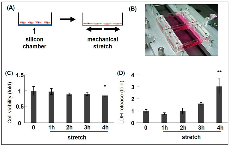
Acute aortic dissection (AAD) and associated ruptures are the leading causes of death in cardiovascular diseases (CVDs). Hypertension is a prime risk factor for AAD. However, the molecular mechanisms underlying AAD remain poorly understood. We previously reported that cyclic mechanical stretch (CMS) leads to the death of rat aortic smooth muscle cells (RASMCs). This review focuses on the mechanisms of CMS-induced vascular smooth muscle cell (VSMC) death. Moreover, we have also discussed the potential therapeutics for preventing AAD and aneurysm ruptures.

Keywords: aortic dissection; cell death; chemokines; hypertension; inducible nitric oxide synthases; mechanical stretch; vascular smooth muscle cell.


Conflict of interest statement

The authors declare no conflicts of interest.

Figures

Figure 1
Figure 1
Cyclic mechanical stretch (CMS) schematic/apparatus and CMS-induced rat aortic smooth muscle cell (RASMC) viability/death data. (A) Schematic describing CMS (mimicking hypertension). (B) The mechanical stretch apparatus (Model STB-1400, STREX Co., Ltd., Osaka, Japan). RASMCs were subjected to CMS (15% elongation) for different time intervals (0–4 h) and incubated for 1 day. Cell death/viability levels were examined using 3-(4,5-dimethylthiazol-2-yl)-2,5-diphenyl-2H-tetrazolium bromide (MTT) (C) and lactate dehydrogenase (LDH) release assays (D). Colorimetric data were normalized to control data by arbitrarily setting their absorbance values (0 h) to 1. Data are indicated by mean ± standard deviation (n = 4). Asterisks refer to significant differences when compared with controls (* p < 0.05, ** p < 0.01). Modified from Zhao et al. [33].
Figure 2
Figure 2
Schematic showing azelnidipine and olmesartan mediated-effects on CMS-induced JNK and p38 activation in RASMCs. AT1R: Angiotensin II receptor type 1; SP600125 (SP): JNK inhibitor; SB203580 (SB): p38 inhibitor; RASMC: rat aortic smooth muscle cell.
Figure 3
Figure 3
CMS-induced-RASMC viability data plus azelnidipine (CS905, CS), olmesartan (Olm), and MAPK inhibitors (SP and SB). RASMCs pre-incubated with Olm, CS, SP, and SB for 20 min were exposed to CMS (15% elongation) for 4 h and incubated for 1 day. Cell viability was examined using MTT assays. Colorimetric analysis of each value was normalized by arbitrarily setting the colorimetric value of the control cells without stretch to 1. Data are indicated by mean ± standard deviation (n = 4). (* p < 0.05 and # p < 0.05 compared with control without stretch and stretch only, respectively). Modified from Zhao et al. [33] and Ito et al. [34].
Figure 4
Figure 4
Candidate gene transcript expression in CMS-exposed RASMCs. RASMCs were exposed to CMS for 4 h after which Cxcl1 (A), Cx3cl1 (B), iNOS (C), NR4A1 (D), Mmp9 (E), and SERPINE1 (F) expression transcripts were evaluated (RT-PCR), expressed as a percentage of control (Ctrl.), and normalized to GAPDH. Data indicate the mean ± standard error (n = 6) * p < 0.05 and ** p < 0.01 versus control. Modified from Zhao et al. [35,36]. NR4A1: nuclear receptor subfamily 4 group A member 1; Mmp9: matrix metalloproteinase-9; SERPINE1: serpin family E member 1; GAPDH: Glyceraldehyde 3-phosphate dehydrogenase.
Figure 5
Figure 5
Graphic showing CMS-induced cell death mechanisms in RASMCs and putative drug targets. CMS applied to VSMCs induces JNK, p38 and STAT1 phosphorylation leading to cell death and evokes down-stream multiple signalling pathways non-specifically. Upon the process, Cxcl1 and Cx3cl1 expression is induced in a JNK-dependent manner, while iNOS expression is induced via STAT1 and p38 signal pathways independently. The induced Cxcl1, Cx3cl1, and iNOS play positive roles in protecting VSMCs from CMS-induced cell death. Nifuroxazide (nif): STAT1 inhibitor; NO: Nitric Oxide; CxcR2: C-X-C motif chemokine receptor 2; Cx3cR1: C-X3-C motif chemokine receptor 1.

Similar articles

Cited by

References

    1. Rodgers J.L., Jones J., Bolleddu S.I., Vanthenapalli S., Rodgers L.E., Shah K., Karia K., Panguluri S.K. Cardiovascular Risks Associated with Gender and Aging. J. Cardiovasc. Dev. Dis. 2019;6:19. doi: 10.3390/jcdd6020019. - DOI - PMC - PubMed
    1. Nedkoff L., Briffa T., Zemedikun D., Herrington S., Wright F.L. Global Trends in Atherosclerotic Cardiovascular Disease. Clin. Ther. 2023;45:1087–1091. doi: 10.1016/j.clinthera.2023.09.020. - DOI - PubMed
    1. Jebari-Benslaiman S., Galicia-García U., Larrea-Sebal A., Olaetxea J.R., Alloza I., Vandenbroeck K., Benito-Vicente A., Martín C. Pathophysiology of Atherosclerosis. Int. J. Mol. Sci. 2022;20:3346. doi: 10.3390/ijms23063346. - DOI - PMC - PubMed
    1. Xiang Q., Tian F., Xu J., Du X., Zhang S., Liu L. New insight into dyslipidemia-induced cellular senescence in atherosclerosis. Biol. Rev. Camb. Philos. Soc. 2022;97:1844–1867. doi: 10.1111/brv.12866. - DOI - PMC - PubMed
    1. Bäck M., Yurdagul A., Jr., Tabas I., Öörni K., Kovanen P.T. Inflammation and its resolution in atherosclerosis: Mediators and therapeutic opportunities. Nat. Rev. Cardiol. 2019;16:389–406. doi: 10.1038/s41569-019-0169-2. - DOI - PMC - PubMed

Grants and funding