. 2024 Feb 23;25(5):2608.
doi: 10.3390/ijms25052608.

Myelin Basic Protein Attenuates Furin-Mediated Bri2 Cleavage and Postpones Its Membrane Trafficking

Affiliations

Myelin Basic Protein Attenuates Furin-Mediated Bri2 Cleavage and Postpones Its Membrane Trafficking

Evgeniya V Smirnova et al. Int J Mol Sci. .

Abstract

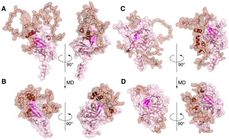
Myelin basic protein (MBP) is the second most abundant protein in the central nervous system and is responsible for structural maintenance of the myelin sheath covering axons. Previously, we showed that MBP has a more proactive role in the oligodendrocyte homeostasis, interacting with membrane-associated proteins, including integral membrane protein 2B (ITM2B or Bri2) that is associated with familial dementias. Here, we report that the molecular dynamics of the in silico-generated MBP-Bri2 complex revealed that MBP covers a significant portion of the Bri2 ectodomain, assumingly trapping the furin cleavage site, while the surface of the BRICHOS domain, which is responsible for the multimerization and activation of the Bri2 high-molecular-weight oligomer chaperone function, remains unmasked. These observations were supported by the co-expression of MBP with Bri2, its mature form, and disease-associated mutants, which showed that in mammalian cells, MBP indeed modulates the post-translational processing of Bri2 by restriction of the furin-catalyzed release of its C-terminal peptide. Moreover, we showed that the co-expression of MBP and Bri2 also leads to an altered cellular localization of Bri2, restricting its membrane trafficking independently of the MBP-mediated suppression of the Bri2 C-terminal peptide release. Further investigations should elucidate if these observations have physiological meaning in terms of Bri2 as a MBP chaperone activated by the MBP-dependent postponement of Bri2 membrane trafficking.

Keywords: artificial intelligence; integral membrane protein 2B (ITM2B/Bri2); intermolecular interaction; myelin basic protein (MBP); protein folding; protein processing regulation.


Conflict of interest statement

The authors declare no conflicts of interest.

Figures

Figure 1
Figure 1
Five AlphaFold2-built 3D models of MBP colored according to their confidence level. pLDDT > 90 (red), pLDDT > 80 (orange), pLDDT > 70 (yellow), pLDDT > 60 (green), pLDDT > 50 (azur), pLDDT > 40 (blue), and pLDDT > 30 (dark blue). Models are numbered (1–5) from maximum to minimum confidence level. Three α-helices and N- and C-termini are indicated.
Figure 2
Figure 2
AlphaFold2-built structures of Bri2, mBri2, disease-associated British and Danish Bri2 variants, and corresponding amyloidogenic peptides. The best models are colored according to their confidence levels: pLDDT > 90 (red), pLDDT > 80 (orange), pLDDT > 70 (yellow), pLDDT > 60 (green), pLDDT > 50 (azur), pLDDT > 40 (blue), and pLDDT > 30 (dark blue). The Arg 244 residues at position P1 in furin cleavage site are shown in spheres, and the Bri23 β-hairpin or its mutated variants are shown in black. N and C indicate the corresponding termini of proteins. The BRICHOS domain and the N-terminal fragment (NTF), which includes the ICD and TM of Bri2, are shown on the right as an overlay of the above structures.
Figure 3
Figure 3
Three complexes with the best interface characteristics obtained by co-folding MBP with various Bri2 fragments using the program AlphaFold-Multimer: the MBP-Bri2 NTF complex 2 (A). MBP-Bri2 complex 2 (B). MBP-Bri2 complex 3 (C). Numbering is given according to Table 1. Two projections (rotated 180 degrees) are shown for each complex. All complexes are prepared in the form of surface cartoon pictograms, where the Bri2 part is colored pink, while Bri23 C-terminal peptide is in magenta, and the MBP part is brown.
Figure 4
Figure 4
The stabilities of the selected structures 1–25 (A) and 5–6 (B) of the modeled MBP-Bri2Δ64 complexes were studied using the 100 ns MD simulation. Time development of the RMSD and Rg throughout the MD trajectory relative to the HADDOCK-built models confirms stabilization of the systems. Residual RMSF values are shown in azur and orange for Bri2 and MBP, respectively.
Figure 5
Figure 5
Molecular dynamics simulation of MBP-Bri2Δ64 complexes results in significant increases in the intermolecular interactions and compacting of the complexes. Two HADDOCK-generated MBP-Bri2 complexes with the best intermolecular interface characteristics: 1–25 (A,B) and 5–6 (C,D) before (A,C) and after (B,D) 100 ns MD simulations. For both experiments, only part of Bri2 molecule (without ITC and with partial TM domain) was used. Before and after MD complexes are shown in two projections (rotated 90 degrees) in the form of surface cartoon pictograms. The Bri2 part is colored pink; the Bri23 peptide is shown in magenta; and the MBP part is colored brown. N- and C-termini of proteins as well as α-helices of MBP are annotated.
Figure 6
Figure 6
Putative structure of MBP-Bri2 complex obtained by the combination of the protein–protein docking and molecular dynamics. MBP is colored brown; Bri2 is colored salmon; and Bri23 peptide is colored pink. Proteolytic processing sites are shown by arrows in the Bri2 structure. Insert shows hydrophobic cavity stabilized by Bri2 R244–MBP Y16 cation–π interaction.
Figure 7
Figure 7
MBP interacts with Bri2 and modulates its intracellular processing. (A) Immunoprecipitation (IP) analysis demonstrating the Bri2-MBP interaction. The IP eluates were separated by SDS-PAGE, transferred to the nitrocellulose membrane, and immunoblotted with indicated antibodies. The plasmids used in transfection are shown on the top. The antibodies used in Western blotting are shown on the right. (B) Schematic representation of Bri2 variants used in the co-expression experiments. (C) Immunoblot of whole-cell lysates (WCLs) of HEK293T cells overexpressing Bri2 variants with MBP (left) or DHFR (right). (D) Quantification of the ratio of non-processed Bri2 (npBri2) in MBP and DHFR co-expressing cells related to WB panel C(i). (E) Quantification of the ratio of npBri2 and its processed form mBri2 related to WB panel (C(i)). (F) Comparison of the protein level of HA-tagged Bri2 and Bri2 KRmut in MBP co-expressing cells and DHFR co-expressing cells related to WB panel (C(ii)). (G) Comparison of the NTF release in MBP co-expressing cells and DHFR co-expressing cells related to WB panel (C(iii)). In (EG), light blue and grey bars indicate MBP and DHFR co-expression, respectively. Average values with standard deviations are shown. Statistically significant difference is marked by asterisk.
Figure 8
Figure 8
Immunofluorescence staining of overexpressed Bri2 KRmut in the presence of MBP or DHFR in HEK 293T cells. Mouse anti-Strep antibodies (BioRad, Hercules, CA, USA) and DyLight649-labeled secondary goat anti-mouse IgG antibodies (Rockland Immunochemicals, Limerick, PA, USA) were used to stain Bri2 proteins. The expressed proteins are annotated on the left.
Figure 9
Figure 9
Bri2 as a potential MBP chaperone. Co-expression of MBP and Bri2 results in suppression of furin, but not ADAM10- and SPPL2a/b-mediated Bri2 processing. Multimerization of BRICHOS domains activates Bri2 high-molecular-weight oligomer chaperone function, which provides MBP membrane trafficking. Cellular organelles are presented in grey, Bri2 and its domains are presented in pink, and MBP is presented in brown.

Similar articles

References

    1. Kim D., Tsai L.-H. Bridging Physiology and Pathology in AD. Cell. 2009;137:997–1000. doi: 10.1016/j.cell.2009.05.042. - DOI - PubMed
    1. Benson M.D., Buxbaum J.N., Eisenberg D.S., Merlini G., Saraiva M.J.M., Sekijima Y., Sipe J.D., Westermark P. Amyloid Nomenclature 2018: Recommendations by the International Society of Amyloidosis (ISA) Nomenclature Committee. Amyloid. 2018;25:215–219. doi: 10.1080/13506129.2018.1549825. - DOI - PubMed
    1. Hardy J.A., Higgins G.A. Alzheimer’s Disease: The Amyloid Cascade Hypothesis. Science. 1992;256:184–185. doi: 10.1126/science.1566067. - DOI - PubMed
    1. Hardy J. The Discovery of Alzheimer-causing Mutations in the APP Gene and the Formulation of the “Amyloid Cascade Hypothesis”. FEBS J. 2017;284:1040–1044. doi: 10.1111/febs.14004. - DOI - PubMed
    1. Alzheimer Disease Genetics Consortium (ADGC) The European Alzheimer’s Disease Initiative (EADI) Cohorts for Heart and Aging Research in Genomic Epidemiology Consortium (CHARGE) Genetic and Environmental Risk in AD/Defining Genetic, Polygenic and Environmental Risk for Alzheimer’s Disease Consortium (GERAD/PERADES) Kunkle B.W., Grenier-Boley B., Sims R., Bis J.C., Damotte V., Naj A.C., et al. Genetic Meta-Analysis of Diagnosed Alzheimer’s Disease Identifies New Risk Loci and Implicates Aβ, Tau, Immunity and Lipid Processing. Nat. Genet. 2019;51:414–430. doi: 10.1038/s41588-019-0358-2. - DOI - PMC - PubMed

LinkOut - more resources