. 2024 Feb 24;25(5):2642.
doi: 10.3390/ijms25052642.

Pulmonary Biodistribution of Platelet-Derived Regenerative Exosomes in a Porcine Model

Affiliations

Pulmonary Biodistribution of Platelet-Derived Regenerative Exosomes in a Porcine Model

Skylar A Rizzo et al. Int J Mol Sci. .

Abstract

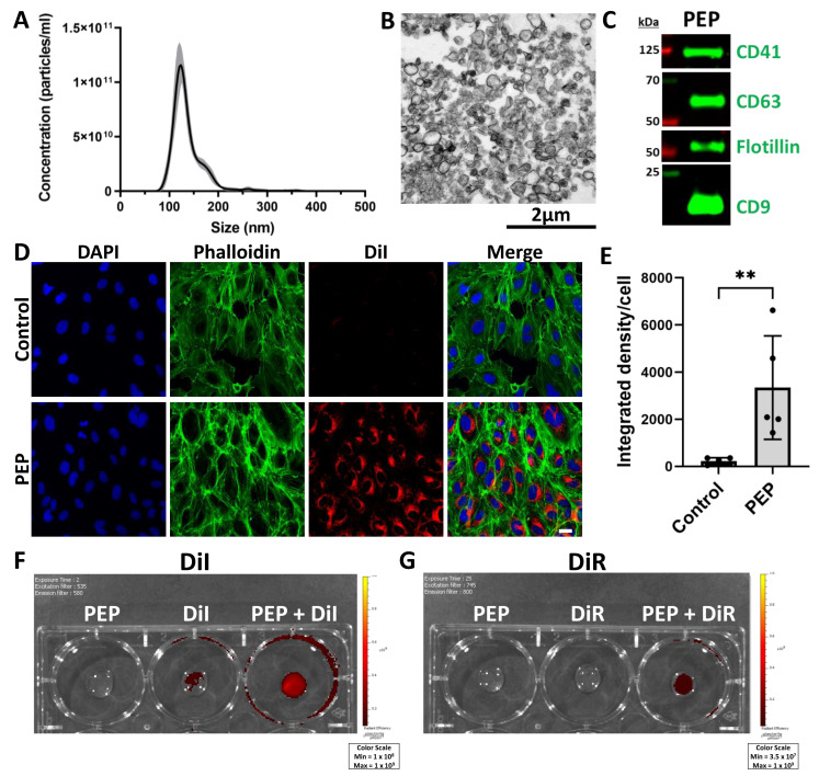
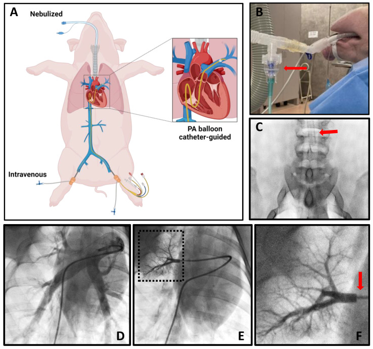
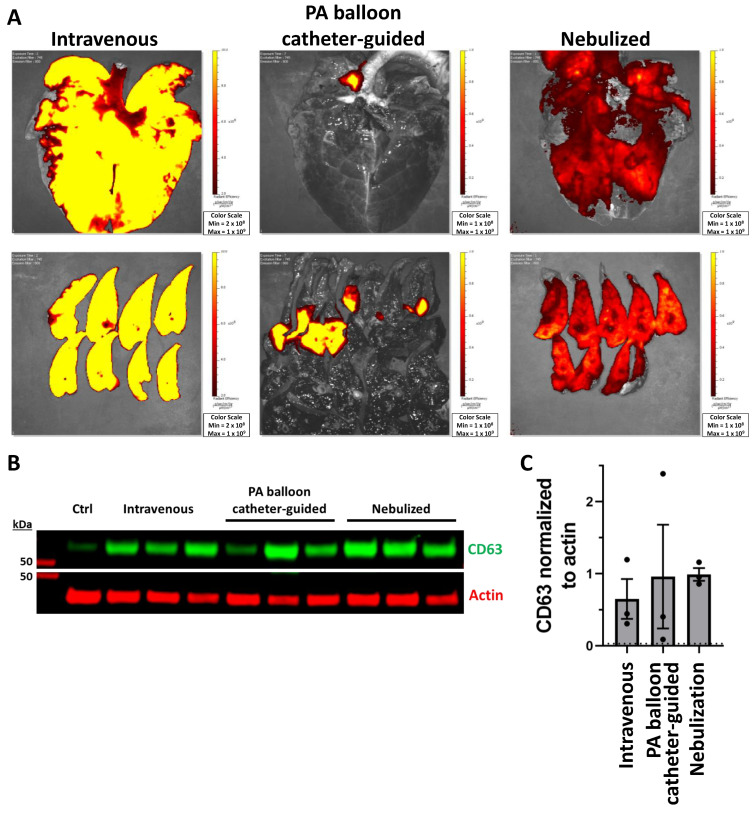
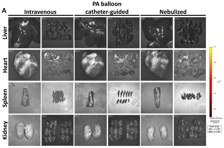
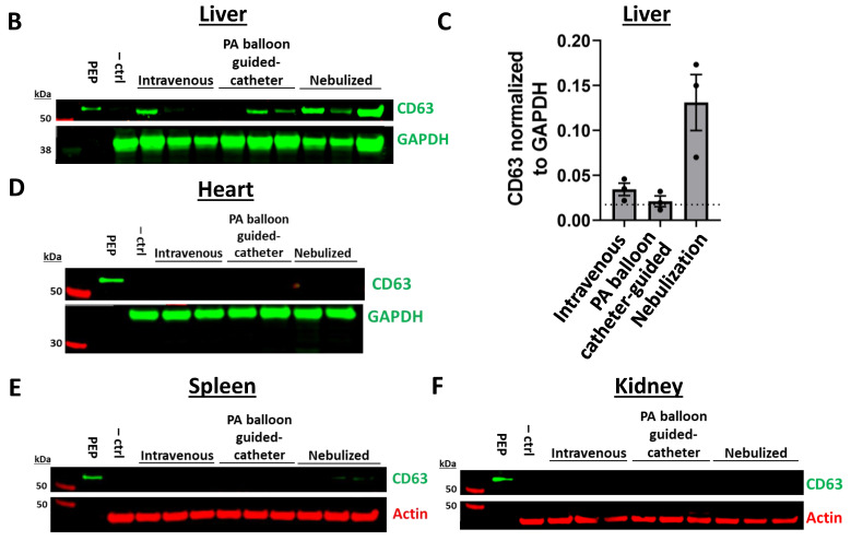
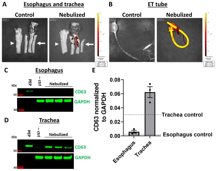
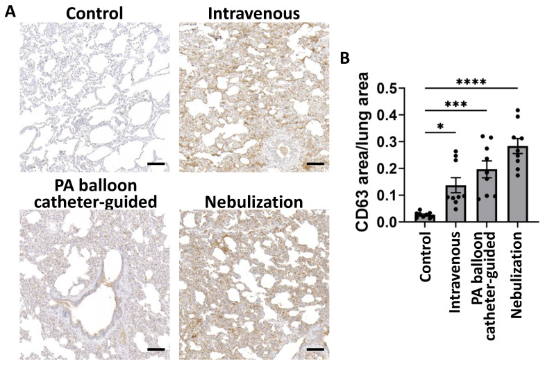
The purpose of this study was to evaluate the biodistribution of a platelet-derived exosome product (PEP), previously shown to promote regeneration in the setting of wound healing, in a porcine model delivered through various approaches. Exosomes were labeled with DiR far-red lipophilic dye to track and quantify exosomes in tissue, following delivery via intravenous, pulmonary artery balloon catheter, or nebulization in sus scrofa domestic pigs. Following euthanasia, far-red dye was detected by Xenogen IVUS imaging, while exosomal protein CD63 was detected by Western blot and immunohistochemistry. Nebulization and intravenous delivery both resulted in global uptake of exosomes within the lung parenchyma. However, nebulization resulted in the greatest degree of exosome uptake. Pulmonary artery balloon catheter-guided delivery provided the further ability to localize pulmonary delivery. No off-target absorption was noted in the heart, spleen, or kidney. However, the liver demonstrated uptake primarily in nebulization-treated animals. Nebulization also resulted in uptake in the trachea, without significant absorption in the esophagus. Overall, this study demonstrated the feasibility of pulmonary delivery of exosomes using nebulization or intravenous infusion to accomplish global delivery or pulmonary artery balloon catheter-guided delivery for localized delivery.

Keywords: biodistribution; exosomes; extracellular vesicles; pulmonary.


Conflict of interest statement

A.B. and P.G.S. and Mayo Clinic hold shares in Rion Inc. but have no other non-financial competing interests. All other authors declare no competing financial or non-financial interests.

Figures

Figure 1
Figure 1
Exosome characterization of PEP and membrane labeling by lipophilic dye. (A) NanoSight analysis of PEP demonstrating distribution of particle size ± standard deviation (gray shading). (n = 3). (B) Transmission electron microscopy image of PEP with scale bar 2 µm. (C) Western blot demonstrating presence of hallmark exosomal proteins CD9, CD63, and Flotillin, as well as platelet integrin CD41. (D) Immunocytochemistry of human umbilical vein endothelial cells following treatment with DiI-labeled PEP or DiI alone (control) in red, counterstained with phalloidin (green) and DAPI (blue), scale bar 20 µm. (E) Quantification (n = 5) of mean integrated density DiI per nuclei (±SD) for Figure 1D. ** p < 0.001 using Mann-Whitney two-tailed t-test. (F) Xenogen image demonstrating fluorescent signal detected in unlabeled PEP, DiI, and PEP + DiI. (G) Xenogen image demonstrating fluorescent signal detected in unlabeled PEP, DiR, and PEP + DiR.
Figure 2
Figure 2
Pulmonary exosome delivery procedures. (A) Cartoon depicting methods employed for pulmonary delivery of exosomes, including nebulization, intravenous, and PA balloon catheter-guided approaches. (B) Photograph demonstrating jet nebulizer (red arrow) attachment to endotracheal tubing. (C) Fluoroscopy image showing placement of endotracheal tube (red arrow) in the trachea above the carina to allow bilateral pulmonary delivery. (D) Pulmonary angiogram of the right lung. (E) Angiogram demonstrating occlusion by balloon catheter of pulmonary artery branch with contrast injection and dashed box indicating area of zoomed in image in Figure 2F. (F) Zoomed-in image from Figure 2E with red arrow indicating area of balloon catheter occlusion.
Figure 3
Figure 3
Pulmonary absorption of exosomes with intravenous, PA balloon catheter-guided, and nebulized delivery. (A) Xenogen imaging of uptake of DiR-labeled PEP in the lungs for intravenous, PA balloon catheter-guided, and nebulized delivery. (B) Western blot demonstrating presence of PEP in control lung (ctrl) tissue compared to lung tissue from intravenous, PA balloon catheter-guided, and nebulized deliveryusing exosomal protein CD63 (green) and loading control actin (red). (C) Quantification (n = 3) of mean fluorescent signal (±SEM) of CD63 normalized to actin loading control; dotted line represents level of control (ctrl).
Figure 4
Figure 4
Off-target absorption of exosomes in the liver, heart, spleen, and kidney with intravenous, PA balloon catheter-guided, and nebulized delivery. (A) Xenogen imaging of uptake of DiR-labeled PEP in the heart, liver, spleen, and kidney with images of entire organ and organ sections shown. (B) Western blots demonstrating presence of PEP in liver tissue compared to PEP and control organ tissue (–ctrl) using exosomal protein CD63 (green) and loading control GAPDH (green). (C) Quantification (n = 3) of mean fluorescent signal (±SEM) of CD63 normalized to GAPDH loading control, with level of control liver tissue shown in dotted line. (DF) Western blots demonstrating presence of PEP in heart (D), spleen (E), and kidney (F) compared to PEP and control organ tissue (–ctrl) using exosomal protein CD63 (green) and loading controls actin (red) or GAPDH (green).
Figure 4
Figure 4
Off-target absorption of exosomes in the liver, heart, spleen, and kidney with intravenous, PA balloon catheter-guided, and nebulized delivery. (A) Xenogen imaging of uptake of DiR-labeled PEP in the heart, liver, spleen, and kidney with images of entire organ and organ sections shown. (B) Western blots demonstrating presence of PEP in liver tissue compared to PEP and control organ tissue (–ctrl) using exosomal protein CD63 (green) and loading control GAPDH (green). (C) Quantification (n = 3) of mean fluorescent signal (±SEM) of CD63 normalized to GAPDH loading control, with level of control liver tissue shown in dotted line. (DF) Western blots demonstrating presence of PEP in heart (D), spleen (E), and kidney (F) compared to PEP and control organ tissue (–ctrl) using exosomal protein CD63 (green) and loading controls actin (red) or GAPDH (green).
Figure 5
Figure 5
Off-target absorption of exosomes in the esophagus and trachea with intravenous, PA balloon catheter-guided, and nebulized delivery. (A) Xenogen imaging of DiR-labeled PEP uptake in the esophagus (white arrowhead) and trachea (white arrow) of control tissue compared to tissue exposed to nebulized PEP (top). (B) Endotracheal (ET) tube demonstrating loss of PEP on plastic tubing also shown (bottom). (C) Western blot demonstrating PEP uptake in the esophagus by nebulization compared to PEP and control pig esophagus tissue (–ctrl) with exosome marker CD63 (green) and GAPDH (green) loading control. (D) Western blot demonstrating PEP uptake in the trachea by nebulization compared to PEP and control pig trachea tissue (–ctrl) with exosome marker CD63 (green) and GAPDH (green) loading control. (E) Quantification (n = 3) of mean fluorescent signal (±SEM) of CD63 normalized to GAPDH loading control for trachea and esophagus with nebulization. The level of control esophagus or trachea tissue is shown with the labeled dotted lines.
Figure 6
Figure 6
Histologic evaluation of pulmonary exosome delivery with intravenous, PA balloon catheter-guided, and nebulized delivery. (A) Immunohistochemical staining of lung tissue with CD63 (brown) with hematoxylin counterstain (blue) with scale bar 100 µm. (B) Mean (±SEM) ratio of CD63 area to total lung tissue area in untreated (control), intravenous, PA balloon catheter-guided, or nebulization delivery methods (n = 9). * p < 0.05, *** p < 0.001, and **** p < 0.0001 using Kruskal–Wallis test.

References

    1. El Andaloussi S., Mäger I., Breakefield X.O., Wood M.J. Extracellular vesicles: Biology and emerging therapeutic opportunities. Nat. Rev. Drug Discov. 2013;12:347–357. doi: 10.1038/nrd3978. - DOI - PubMed
    1. Cheng L., Hill A.F. Therapeutically harnessing extracellular vesicles. Nat. Rev. Drug Discov. 2022;21:379–399. doi: 10.1038/s41573-022-00410-w. - DOI - PubMed
    1. Wiklander O.P., Nordin J.Z., O’Loughlin A., Gustafsson Y., Corso G., Mager I., Vader P., Lee Y., Sork H., Seow Y., et al. Extracellular vesicle in vivo biodistribution is determined by cell source, route of administration and targeting. J. Extracell. Vesicles. 2015;4:26316. doi: 10.3402/jev.v4.26316. - DOI - PMC - PubMed
    1. Choi H., Choi Y., Yim H.Y., Mirzaaghasi A., Yoo J.K., Choi C. Biodistribution of Exosomes and Engineering Strategies for Targeted Delivery of Therapeutic Exosomes. Tissue Eng. Regen. Med. 2021;18:499–511. doi: 10.1007/s13770-021-00361-0. - DOI - PMC - PubMed
    1. Zhai Z., Cui T., Chen J., Mao X., Zhang T. Advancements in engineered mesenchymal stem cell exosomes for chronic lung disease treatment. J. Transl. Med. 2023;21:895. doi: 10.1186/s12967-023-04729-9. - DOI - PMC - PubMed

LinkOut - more resources