. 2024 Feb 24;25(5):2643.
doi: 10.3390/ijms25052643.

HLA-G Expression/Secretion and T-Cell Cytotoxicity in Missed Abortion in Comparison to Normal Pregnancy

Affiliations

HLA-G Expression/Secretion and T-Cell Cytotoxicity in Missed Abortion in Comparison to Normal Pregnancy

Antonia Terzieva et al. Int J Mol Sci. .

Abstract

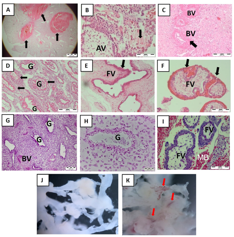
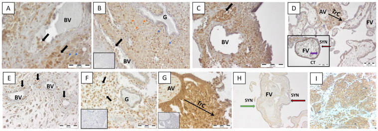
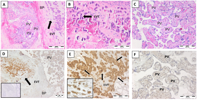
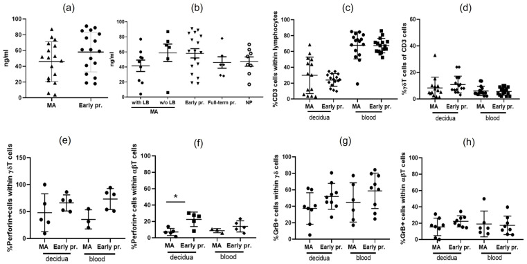
The main role of HLA-G is to protect the semi-allogeneic embryo from immune rejection by proper interaction with its cognate receptors on the maternal immune cells. Spontaneous abortion is the most common adverse pregnancy outcome, with an incidence rate between 10% and 15%, with immunologic dysregulation being thought to play a role in some of the cases. In this study, we aimed to detect the membrane and soluble HLA-G molecule at the maternal-fetal interface (MFI) and in the serum of women experiencing missed abortion (asymptomatic early pregnancy loss) in comparison to the women experiencing normal early pregnancy. In addition, the proportion of T cells and their cytotoxic profile was evaluated. We observed no difference in the spatial expression of HLA-G at the MFI and in its serum levels between the women with missed abortions and those with normal early pregnancy. In addition, comparable numbers of peripheral blood and decidual total T and γδT cells were found. In addition, as novel data we showed that missed abortion is not associated with altered extravilous invasion into uterine blood vessels and increased cytotoxicity of γδT cells. A strong signal for HLA-G on non-migrating extravilous trophoblast in the full-term normal placental bed was detected. In conclusion, HLA-G production at the MFI or in the blood of the women could not be used as a marker for normal pregnancy or missed abortions.

Keywords: HLA-G; T cells; missed abortion; γδ T cells.


Conflict of interest statement

The authors declare no conflicts of interest. The funders had no role in the design of the study; in the collection, analyses, or interpretation of data; in the writing of the manuscript, or in the decision to publish the results.

Figures

Figure 1
Figure 1
Maternal–fetal interface and explanted placental villi in MA (AF,J) and normal early pregnancy (GI,K). (AF): MA decidua with massive blood cloths (black arrow, A) and anchoring villa and blood vessels with necrotic tissue (black arrow, (B) and (C), respectively). Note the destroyed decidual glands with massive immune infiltration around (D). The placental villi are immature, inflamed, and irregular in shapes (black arrow, E), showing numerous clots (black arrow, F). Representative images for n = 10. Explanted placental villi derived from aborted pregnancy (J, n = 3) are not vital, and they are shown by a lack of blood vessels which are numerous in explanted normal placental villi (red arrows, K). (GI): Healthy decidua with normal glands and blood vessels (G,H) as well as floating mature placental villi bathed in the maternal blood into intervillous spaces (I). Representative images for n = 10. Hematoxylin and eosin staining. BVs—blood vessels, G—gland, AV—anchoring villa, FV—floating villi, MB—maternal blood. Scale bar represents 200 μm (A,C,F,G,H) and 100 μm (B,D,I). Magnification 4× (J,K) and 10× (E).
Figure 2
Figure 2
In situ expression of non-classical HLA-G molecule at MFI in MA (AD) and normal early pregnancy (EH). Representative images of n = 10 for each group. Note the similar pattern in HLA-G expression in decidua and placental villi (anchoring and floating) between women with MA and normal pregnancy. HLA-G+ EVT invading the decidua and blood vessels (black arrow, A,B,E) but not the decidual glands (B,F). Endovascular plugs of HLA-G+ EVT in aborted decidua (black arrow, C). HLA-G+ immune cells (orange arrow) as well as some weakly positive signals for HLA-G DSC (blue arrows) were observed (A,B). Trophoblast columns (TrC) of the anchoring villa contain intermediate cytotrophoblasts positive for HLA-G (D,G). The syntitiotrophoblast of the floating villi do not express HLA-G (red arrow, D,H) unlike underlying cytotrophoblasts (purple arrow, D). (I)—HLA-G-positive breast cancer tissue serving as positive control for the specificity of the staining. Negative control sections are given as insets in (B,F,G), and the inset in (D) shows floating villa at higher magnification. BVs—blood vessels, G—gland, AV—anchoring villa, FV—floating villi, syn—syntitiotrophoblasts, CTs—cytotrophoblasts, TrC—trophoblast column. Scale bar represents 200 μm (A,C,D), 180 μm (I), and 100 μm (E,F,G). Magnification 10× (B) and 20× (H). The scale bars of the insets correspond to those of the main images, except for (D) where the inset scale bar is 100 μm.
Figure 3
Figure 3
The presence of EVT cells in the basal plate of full-term placenta. (A)—full-term placenta and EVT cells (black arrow) in its basal plate; (B)—EVT at higher magnification; (C)—placental villi; (D)—HLA-G-positive EVT in the basal plate of the placenta (black arrow); (E)—HLA-G+ EVT cells (black arrows) at higher magnification; the inset shows round-shape stationary EVTs with endoduplication; (F)—HLA-G-negative placental villi. (AC)—hematoxylin and eosin staining. (DF)—detection of HLA-G; an inset of (D) is a negative control staining. PV—placental villi, BP—basal plate, EVTs—extravillous trophoblasts. Representative images of n = 5. Scale bar represents 200 μm (A,C,D) and 100 μm (B,E,F). The scale bar of the inset of (D) correspond to that of the main image, the scale bar of the inset of (E) is 20 μm.
Figure 4
Figure 4
Serum sHLA-G levels, T-cell, and cytotoxic T-cell proportions of women with MA and normal pregnancy. No significant difference in the serum levels of sHLA-G was detected between women experiencing MA and those experiencing normal pregnancy (a) as well as between women experiencing MA with or without live birth, with normal early or late pregnancy and non-pregnant, non-laboring women (b); (c) T-cell number in the blood and decidua of women experiencing MA and normal pregnancy; (d) γδT-cell number in the blood and decidua of women experiencing MA and normal pregnancy; (e) γδT cells positive for perforin in the blood and decidua of women experiencing MA and normal pregnancy; (f) αβT cells positive for perforin in the blood and decidua of women experiencing MA and normal pregnancy; (g) γδT cells positive for granzyme B in the blood and decidua of women experiencing MA and normal pregnancy; (h) αβT cells positive for granzyme B in the blood and decidua of women experiencing MA and normal pregnancy. MA—missed abortion, LB—live birth, NP—non-pregnant. Mann–Whitney test, GraphPad Prism v8, * p < 0.05.

Similar articles

Cited by

References

    1. Meeusen E.N., Bischof R.J., Lee C. Comparative T-cell responses during pregnancy in large animals and humans. Am. J. Reprod. Immunol. 2001;46:169–179. doi: 10.1111/j.8755-8920.2001.460208.x. - DOI - PubMed
    1. Lala P., Graham C. Mechanisms of trophoblast invasiveness and their control: The role of proteases and protease inhibitors. Cancer Metastasis Rev. 1990;9:369–379. doi: 10.1007/BF00049525. - DOI - PubMed
    1. Aluvihare V.R., Kallikourdis M., Betz A.G. Regulatory T cells mediate maternal tolerance to the fetus. Nat. Immunol. 2004;5:266–271. doi: 10.1038/ni1037. - DOI - PubMed
    1. Hanna J., Goldman-Wohl D., Hamani Y., Avraham I., Greenfield C., Natanson-Yaron S., Prus D., Cohen-Daniel L., I Arnon T., Manaster I., et al. Decidual NK cells regulate key developmental processes at the human fetalmaternal interface. Nat. Med. 2006;12:1065–1074. doi: 10.1038/nm1452. - DOI - PubMed
    1. Vento-Tormo R., Efremova M., Botting R.A., Turco M.Y., Vento-Tormo M., Meyer K.B., Park J.E., Stephenson E., Polański K., Goncalves A., et al. Single-cell reconstruction of the early maternal-fetal interface in humans. Nature. 2018;563:347–353. doi: 10.1038/s41586-018-0698-6. - DOI - PMC - PubMed