A Systematic Review and Meta-Analysis of the Pathology Underlying Aneurysm Enhancement on Vessel Wall Imaging
- PMID: 38473947
- PMCID: PMC10931983
- DOI: 10.3390/ijms25052700
A Systematic Review and Meta-Analysis of the Pathology Underlying Aneurysm Enhancement on Vessel Wall Imaging
Abstract
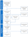
Intracranial aneurysms are common, but only a minority rupture and cause subarachnoid haemorrhage, presenting a dilemma regarding which to treat. Vessel wall imaging (VWI) is a contrast-enhanced magnetic resonance imaging (MRI) technique used to identify unstable aneurysms. The pathological basis of MR enhancement of aneurysms is the subject of debate. This review synthesises the literature to determine the pathological basis of VWI enhancement. PubMed and Embase searches were performed for studies reporting VWI of intracranial aneurysms and their correlated histological analysis. The risk of bias was assessed. Calculations of interdependence, univariate and multivariate analysis were performed. Of 228 publications identified, 7 met the eligibility criteria. Individual aneurysm data were extracted for 72 out of a total of 81 aneurysms. Univariate analysis showed macrophage markers (CD68 and MPO, p = 0.001 and p = 0.002), endothelial cell markers (CD34 and CD31, p = 0.007 and p = 0.003), glycans (Alcian blue, p = 0.003) and wall thickness (p = 0.030) were positively associated with enhancement. Aneurysm enhancement therefore appears to be associated with inflammatory infiltrate and neovascularisation. However, all these markers are correlated with each other, and the literature is limited in terms of the numbers of aneurysms analysed and the parameters considered. The data are therefore insufficient to determine if these associations are independent of each other or of aneurysm size, wall thickness and rupture status. Thus, the cause of aneurysm-wall enhancement currently remains unknown.
Keywords: aneurysm-wall enhancement; histology; intracranial aneurysm; magnetic resonance imaging; pathology; vessel wall imaging.
Conflict of interest statement
The authors declare no conflicts of interest.
Figures


References
Publication types
MeSH terms
LinkOut - more resources
Full Text Sources
Medical
Research Materials
Miscellaneous

