. 2024 Feb 27;25(5):2732.
doi: 10.3390/ijms25052732.

Neurotrophins and Trk Neurotrophin Receptors in the Retina of Adult Killifish (Nothobranchius guentheri)

Affiliations

Neurotrophins and Trk Neurotrophin Receptors in the Retina of Adult Killifish (Nothobranchius guentheri)

Caterina Porcino et al. Int J Mol Sci. .

Abstract

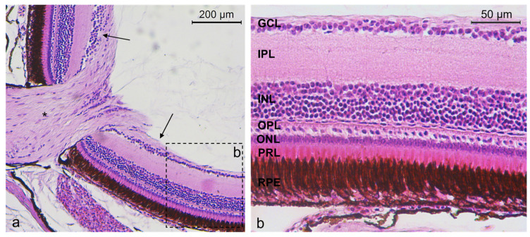
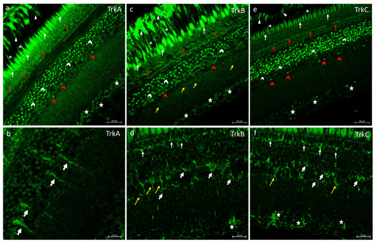
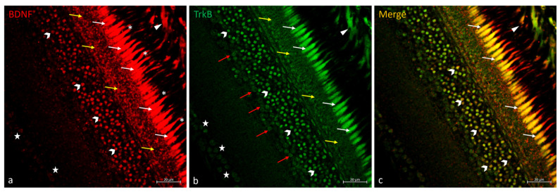
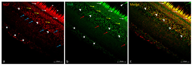
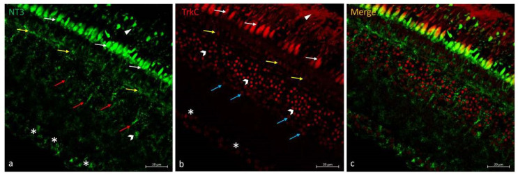
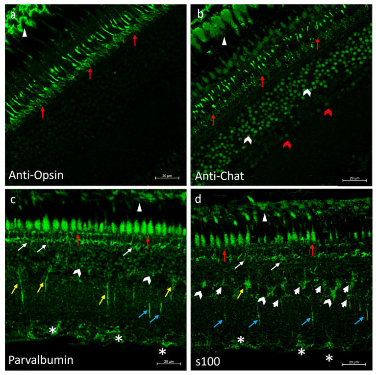
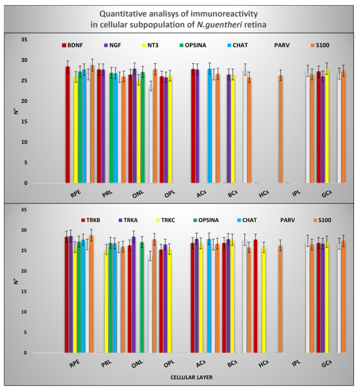
Specific subpopulations of neurons in nerve and sensory systems must be developed and maintained, and this is accomplished in significant part by neurotrophins (NTs) and the signaling receptors on which they act, called tyrosine protein kinase receptors (Trks). The neurotrophins-tyrosine protein kinase receptors (NTs/Trks) system is involved in sensory organ regulation, including the visual system. An NTs/Trks system alteration is associated with neurodegeneration related to aging and diseases, including retinal pathologies. An emergent model in the field of translational medicine, for instance, in aging study, is the annual killifish belonging to the Nothobranchius genus, thanks to its short lifespan. Members of this genus, such as Nothobranchius guentheri, and humans share a similar retinal stratigraphy. Nevertheless, according to the authors' knowledge, the occurrence and distribution of the NTs/Trks system in the retina of N. guentheri has never been investigated before. Therefore, the present study aimed to localize neurotrophin BDNF, NGF, and NT-3 and TrkA, TrkB, and TrkC receptors in the N. guentheri retina using the immunofluorescence method. The present investigation demonstrates, for the first time, the occurrence of the NTs/Trks system in N. guentheri retina and, consequently, the potential key role of these proteins in the biology and survival of the retinal cells.

Keywords: N. guentheri; Trks; neurotrophins; retina; translational medicine.


Conflict of interest statement

The authors declare no conflicts of interest.

Figures

Figure 1
Figure 1
(a) N. guentheri retina (arrows), optic nerve (asterisk). (b) Stratigraphy of N. guentheri retina: RPE, PRL, ONL, OPL, INL, IPL, GCL. Hematoxylin-Eosin. Magnification (a) 20× (b) 40×.
Figure 2
Figure 2
Trks immunostaining in N. guentheri retina. (a) TrkA-immunoreactivity in the cytoplasmic prolongations of the cells of the RPE (arrowheads); in the inner segment (arrows) and outer segment (asterisk) of the photoreceptors layer; in the OPL (red arrows); in different subpopulation of ACs (white and red gallon-arrows); in the soma of GCs (stars). (b) BCs TrkA immunopositive (white bold arrows). (c) TrkB immunoreactivity in the cytoplasmic prolongations of the cells of the RPE (arrowheads); in the inner segment (arrows) and outer segment (asterisks) of the photoreceptors layer; in the OPL (red arrows); in several subpopulation of ACs (white and red gallon-arrows); in cellular prolongation in IPL (yellow arrows); in the soma of GCs (stars). (d) TrkB immunostaining in the inner segment of PRL (arrows indicate the soma of rods and cones); in BCs (yellow arrows); in the HCs (white bold cells), and in GCs (stars). (e) TrkC immunoreactivity in the cytoplasmic prolongations of the cells of the RPE (arrowheads); in the inner segment (arrows) and outer segment (asterisk) of the photoreceptors layer; in the OPL (red arrows); in the INL several subpopulations of ACs (white and red gallon-arrows) and, in the soma of GCs (stars). (f) TrkC immunostaining in the inner segment of PRL (arrows indicate the soma of rods and cones); in the BCs (yellow arrows); in the HCs (white bold cells), and in the GCs (stars). (ac) Magnification 40×; Scale bar 20 µm. (df) Magnification 63×; scale bar 10 µm.
Figure 3
Figure 3
BDNF/TrkB immunostaining in N. guentheri retina. (a) BDNF-immunoreactivity in the cytoplasmic prolongations of the cells of the RPE (arrowheads); in the inner segment (white-arrows) and outer segment of PRL (asterisk); in the OPL (yellow-arrows); in a subpopulation of ACs (gallon-arrows); weakly stained in the soma of GCs (stars). (b) TrkB-immunoreactivity in the cytoplasmic prolongations of the cells of the RPE (arrowheads); in the inner segment of PRL (white-arrows); in the OPL (yellow-arrows); in different subpopulations of ACs (gallon-arrows and red-arrows); in the soma of GCs (stars) poorly stained. (c) Colocalization view of BDNF and TrkB in the cytoplasmic prolongations of the cells of the RPE (arrowheads); in the inner segment of PRL (white-arrows); in a subpopulation of ACs (gallon-arrows). Some ACs and GCs TrkB immunopositive did not show double staining with BDNF. A similar condition was observed for the OPL: BDNF and TrkB immunostained but non-overlapping. Magnification 40×. Scale bar 20 µm.
Figure 4
Figure 4
NGF/TrkA immunostaining in N. guentheri retina. (a) NGF-immunoreactivity in the inner segment (white-arrows) and outer segment (asterisk) of the photoreceptors layer; in the OPL (yellow arrows); in a subpopulation of ACs (gallon-arrows); in the BCs (blue arrows); in the soma of GCs (stars). (b) TrkA-immunoreactivity in the cytoplasmic prolongations of the RPE (arrowheads); in the inner segment of PRL (white-arrows); in the OPL (yellow-arrows); in different subpopulation of ACs (gallon-arrows and red-arrows); in the soma of GCs (stars) poorly stained. (c) Colocalization view of NGF and TrkA in the inner segment of PRL (white-arrows) and in a subpopulation of ACs (gallon-arrows). Some ACs and GCs TrkA immunopositive did not show double staining with NGF. A similar condition was observed for the OPL: NGF and TrkA immunostained but non-overlapping. Magnification 40×. Scale bar 20 µm.
Figure 5
Figure 5
NT-3/TrkC immunostaining in N. guentheri retina. (a) NT-3-immunoreactivity in the cytoplasmic prolongations of the cells of the RPE (arrowheads); in the inner segment of PRL (white arrows); in OPL (yellow-arrows); in the BCs (red arrows); in the soma of GCs (asterisks). (b) TrkC-immunoreactivity in the cytoplasmic prolongations of the cells of the RPE (arrowheads); in the outer segment of PRL (white-arrows), in the OPL (yellow-arrows); in different subpopulations of ACs (gallon-arrows and blue-arrows); in the soma of GCs (asterisks) poorly stained. (c) No colocalization view was observed. Magnification 40×. Scale bar 20 µm.
Figure 6
Figure 6
Anti-opsin, anti-chat, parvalbumin, and s100 as specific markers of subpopulation cells in N. guentheri retina. (a) Anti-Opsin immunoreactivity in the cytoplasmatic prolongation of RPE (arrowheads) and in the rods (red arrows) of PRL. (b) Anti-chat immunoreactivity in the cytoplasmatic prolongation of RPE (arrowheads); in the outer segment of the photoreceptor layer (red arrows); in different subpopulations of ACs (white and red gallon-arrows). (c) Parvalbumin immunoreactivity in the cytoplasmatic prolongation of RPE (arrowheads); in the outer segment (red arrows) and inner segment (white arrows) of PRL; in the ACs (gallon-arrows); in the BCs (yellow arrows) of INL; in the cellular prolongation in IPL (blue arrows); in the soma of GCs (asterisk). (d) s100p immunoreactivity in the cytoplasmatic prolongation of RPE (arrowheads); in the outer segment (red arrows) and inner segment (white arrows) in PRL; in the BCs (yellow arrows), HCs (white bold arrows), ACs (gallon arrows) in the INL; in cellular prolongation of the IPL (blue arrows); in the soma of GCs (asterisk). Magnification 40×. Scale bar 20 µm.
Figure 7
Figure 7
Graphical representation of immunoreactivity quantitative analysis in: RPE, PRL (inner and outer segments), OPL, ACs, BCs, HCs, IPL, and GCs detected by BDNF, NGF, NT-3, TrkA, TrkB, TrkC, anti-Opsin, anti-Chat, Parvalbumin and, s100p. The statistical analysis shows a different distribution pattern of the antibodies used in this study in cellular layer of N. guentheri retina. N°: mean of retinal layer cells immunopositive to BDNF, NGF, NT-3, TrkA, TrkB, TrkC, anti-Opsin, anti-Chat, Parvalbumin and, s100p.

Similar articles

Cited by

References

    1. Nikiforov-Nikishin D.L., Irkha V.A., Kochetkov N.I., Kalita T.L., Nikiforov-Nikishin A.L., Blokhin E.E., Antipov S.S., Makarenkov D.A., Zhavnerov A.N., Glebova I.A., et al. Some Aspects of Development and Histological Structure of the Visual System of Nothobranchius guentheri. Animals. 2021;11:2755. doi: 10.3390/ani11092755. - DOI - PMC - PubMed
    1. Ostrander G.K., Hopkins J. The Laboratory Fish. Elsevier; Amsterdam, The Netherlands: 2000.
    1. Catania S., Germana A., Cabo R., Ochoa-Erena F., Guerrera M., Hannestad J., Represa J., Vega J. Neurotrophin and Trk neurotrophin receptors in the inner ear of Salmo salar and Salmo trutta. J. Anat. 2007;210:78–88. doi: 10.1111/j.1469-7580.2006.00673.x. - DOI - PMC - PubMed
    1. García-Suárez O., Germanà A., Hannestad J., Pérez-Pérez M., Esteban I., Naves F.J., Vega J.A. Changes in the expression of the nerve growth factor receptors TrkA and p75LNGR in the rat thymus with ageing and increased nerve growth factor plasma levels. Cell Tissue Res. 2000;301:225–234. doi: 10.1007/s004419900133. - DOI - PubMed
    1. García-Suárez O., Pérez-Pérez M., Germanà A., Esteban I., Germanà G. Involvement of growth factors in thymic involution. Microsc. Res. Tech. 2003;62:514–523. doi: 10.1002/jemt.10413. - DOI - PubMed

Supplementary concepts