

Is Silver Addition to Scaffolds Based on Polycaprolactone Blended with Calcium Phosphates Able to Inhibit Candida albicans and Candida auris Adhesion and Biofilm Formation?
- PMID: 38474027
- PMCID: PMC10931636
- DOI: 10.3390/ijms25052784
Is Silver Addition to Scaffolds Based on Polycaprolactone Blended with Calcium Phosphates Able to Inhibit Candida albicans and Candida auris Adhesion and Biofilm Formation?
Abstract
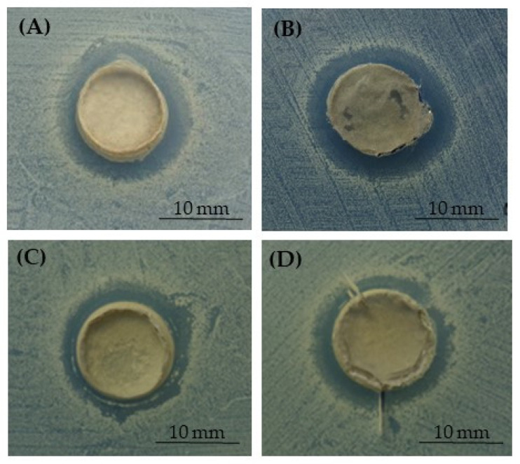
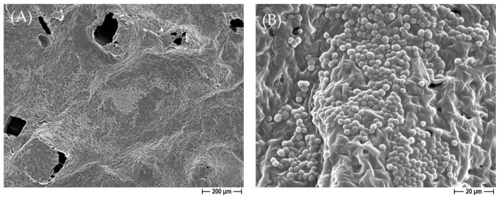
Candida spp. periprosthetic joint infections are rare but difficult-to-treat events, with a slow onset, unspecific symptoms or signs, and a significant relapse risk. Treatment with antifungals meets with little success, whereas prosthesis removal improves the outcome. In fact, Candida spp. adhere to orthopedic devices and grow forming biofilms that contribute to the persistence of this infection and relapse, and there is insufficient evidence that the use of antifungals has additional benefits for anti-biofilm activity. To date, studies on the direct antifungal activity of silver against Candida spp. are still scanty. Additionally, polycaprolactone (PCL), either pure or blended with calcium phosphate, could be a good candidate for the design of 3D scaffolds as engineered bone graft substitutes. Thus, the present research aimed to assess the antifungal and anti-biofilm activity of PCL-based constructs by the addition of antimicrobials, for instance, silver, against C. albicans and C. auris. The appearance of an inhibition halo around silver-functionalized PCL scaffolds for both C. albicans and C. auris was revealed, and a significant decrease in both adherent and planktonic yeasts further demonstrated the release of Ag+ from the 3D constructs. Due to the combined antifungal, osteoproliferative, and biodegradable properties, PCL-based 3D scaffolds enriched with silver showed good potential for bone tissue engineering and offer a promising strategy as an ideal anti-adhesive and anti-biofilm tool for the reduction in prosthetic joints of infections caused by Candida spp. by using antimicrobial molecule-targeted delivery.
Keywords: Candida albicans; Candida auris; Saos-2 cells’ osteogenic differentiation; anti-adhesive/antifungal properties; biofilm inhibition; calcium phosphates; poly(ε-caprolactone)-based biomaterial; silver.
Conflict of interest statement
The authors declare no conflicts of interest.
Figures


Similar articles
-
Nanoparticles in the battle against Candida auris biofilms: current advances and future prospects.Drug Deliv Transl Res. 2025 May;15(5):1496-1512. doi: 10.1007/s13346-024-01749-w. Epub 2024 Nov 26. Drug Deliv Transl Res. 2025. PMID: 39589626 Free PMC article. Review.
-
New Generation of Osteoinductive and Antimicrobial Polycaprolactone-Based Scaffolds in Bone Tissue Engineering: A Review.Polymers (Basel). 2024 Jun 12;16(12):1668. doi: 10.3390/polym16121668. Polymers (Basel). 2024. PMID: 38932017 Free PMC article. Review.
-
Tuning of Silver Content on the Antibacterial and Biological Properties of Poly(ɛ-caprolactone)/Biphasic Calcium Phosphate 3D-Scaffolds for Bone Tissue Engineering.Polymers (Basel). 2023 Aug 31;15(17):3618. doi: 10.3390/polym15173618. Polymers (Basel). 2023. PMID: 37688244 Free PMC article.
-
The Emerging Pathogen Candida auris: Growth Phenotype, Virulence Factors, Activity of Antifungals, and Effect of SCY-078, a Novel Glucan Synthesis Inhibitor, on Growth Morphology and Biofilm Formation.Antimicrob Agents Chemother. 2017 Apr 24;61(5):e02396-16. doi: 10.1128/AAC.02396-16. Print 2017 May. Antimicrob Agents Chemother. 2017. PMID: 28223375 Free PMC article.
-
Novel Silver-Functionalized Poly(ε-Caprolactone)/Biphasic Calcium Phosphate Scaffolds Designed to Counteract Post-Surgical Infections in Orthopedic Applications.Int J Mol Sci. 2021 Sep 21;22(18):10176. doi: 10.3390/ijms221810176. Int J Mol Sci. 2021. PMID: 34576339 Free PMC article.
Cited by
-
Nanoparticles in the battle against Candida auris biofilms: current advances and future prospects.Drug Deliv Transl Res. 2025 May;15(5):1496-1512. doi: 10.1007/s13346-024-01749-w. Epub 2024 Nov 26. Drug Deliv Transl Res. 2025. PMID: 39589626 Free PMC article. Review.
-
New Generation of Osteoinductive and Antimicrobial Polycaprolactone-Based Scaffolds in Bone Tissue Engineering: A Review.Polymers (Basel). 2024 Jun 12;16(12):1668. doi: 10.3390/polym16121668. Polymers (Basel). 2024. PMID: 38932017 Free PMC article. Review.
References
-
- Florea D.A., Grumezescu V., Bîrcă A.C., Vasile B., Ștefan, Mușat M., Chircov C., Stan M.S., Grumezescu A.M., Andronescu E., et al. Design, Characterization, and Antibacterial Performance of MAPLE-Deposited Coatings of Magnesium Phosphate-Containing Silver Nanoparticles in Biocompatible Concentrations. Int. J. Mol. Sci. 2022;23:7910. doi: 10.3390/ijms23147910. - DOI - PMC - PubMed
-
- Holešová S., Čech Barabaszová K., Hundáková M., Ščuková M., Hrabovská K., Joszko K., Antonowicz M., Gzik-Zroska B. Development of Novel Thin Polycaprolactone (PCL)/Clay Nanocomposite Films with Antimicrobial Activity Promoted by the Study of Mechanical, Thermal, and Surface Properties. Polymers. 2021;13:3193. doi: 10.3390/polym13183193. - DOI - PMC - PubMed
MeSH terms
Substances
Grants and funding
LinkOut - more resources
Full Text Sources
Medical
Miscellaneous
