Review
. 2024 Feb 28;25(5):2782.
doi: 10.3390/ijms25052782.

Similarities in Structure and Function of UDP-Glycosyltransferase Homologs from Human and Plants

Affiliations
Review

Similarities in Structure and Function of UDP-Glycosyltransferase Homologs from Human and Plants

Mary Caroline L Lethe et al. Int J Mol Sci. .

Abstract

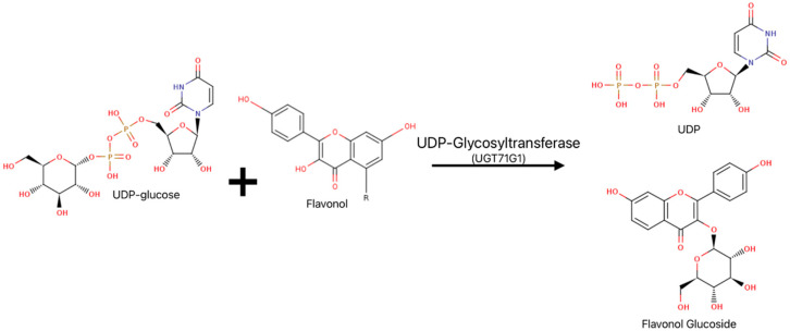
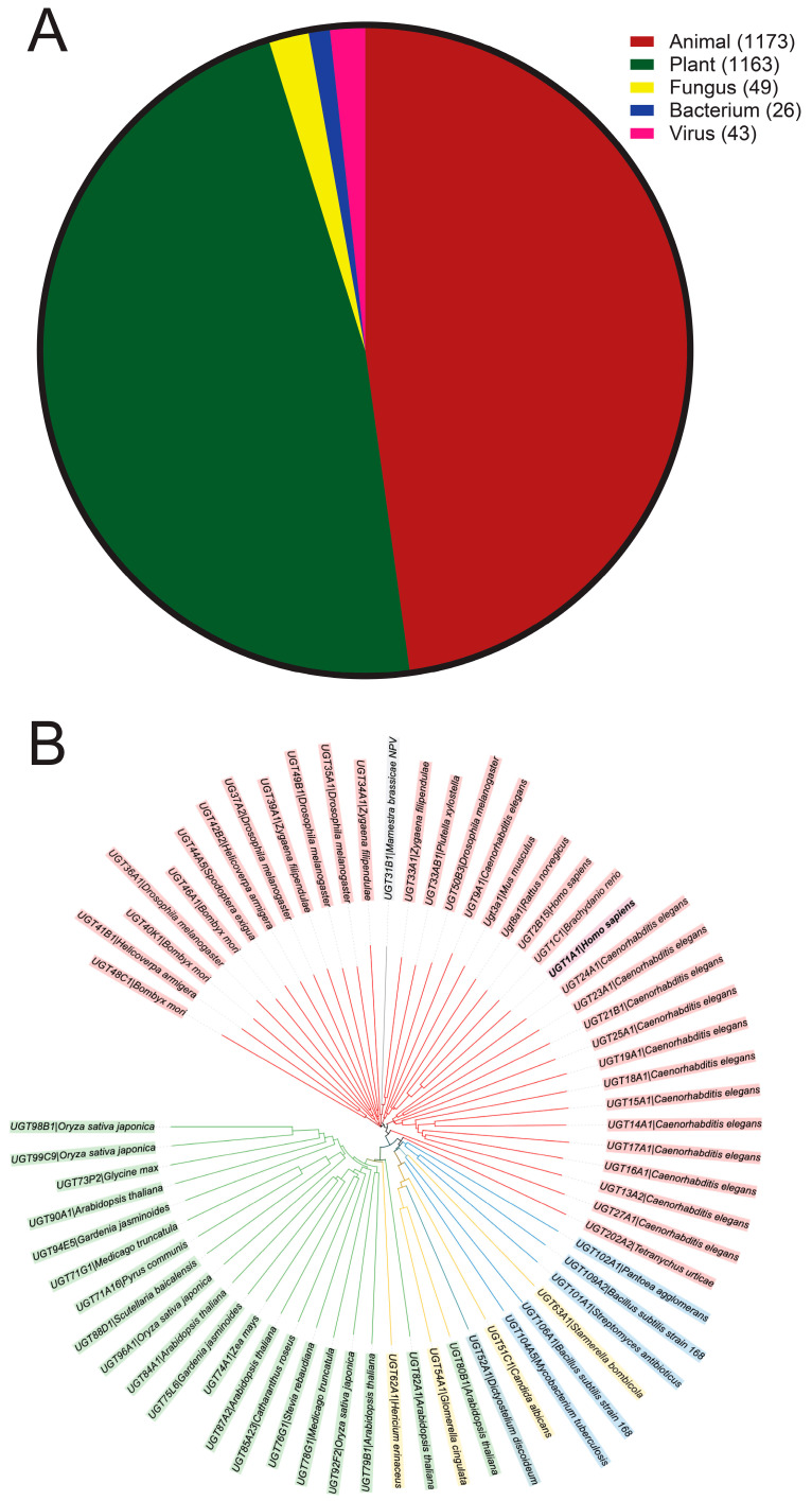
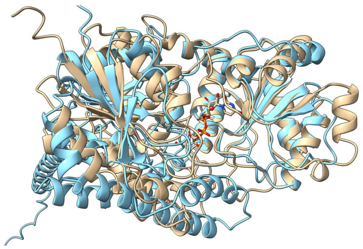
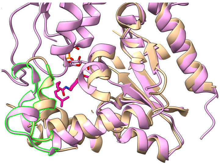
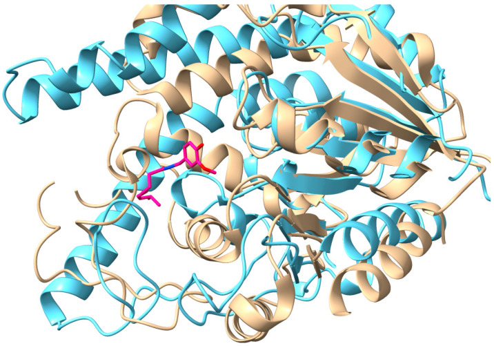
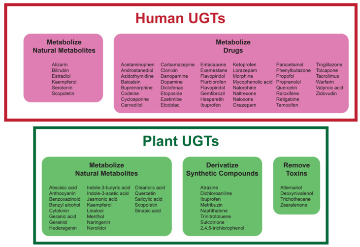
The uridine diphosphate glycosyltransferase (UGT) superfamily plays a key role in the metabolism of xenobiotics and metabolic wastes, which is essential for detoxifying those species. Over the last several decades, a huge effort has been put into studying human and mammalian UGT homologs, but family members in other organisms have been explored much less. Potentially, other UGT homologs can have desirable substrate specificity and biological activities that can be harnessed for detoxification in various medical settings. In this review article, we take a plant UGT homology, UGT71G1, and compare its structural and biochemical properties with the human homologs. These comparisons suggest that even though mammalian and plant UGTs are functional in different environments, they may support similar biochemical activities based on their protein structure and function. The known biological functions of these homologs are discussed so as to provide insights into the use of UGT homologs from other organisms for addressing human diseases related to UGTs.

Keywords: UGT-related diseases; glycosylation; substrate specificity; uridine diphosphate glycosyltransferases.


Conflict of interest statement

The authors declare no conflicts of interest.

Figures

Figure 1
Figure 1
General UGT reaction scheme represented by UGT71G1. Among plant UGT homologs, UDP-glucose is the preferred UDP sugar donor; flavonols are natural plant metabolites that serve as acceptors. Functional groups that involve nitrogen are shown in blue and those with oxygen are shown in red.
Figure 2
Figure 2
UGT homologs from different kingdoms. (A) Identified UGT homologs from different kingdoms. Information was collected from the database of the UGT Nomenclature Committee. The number of UGT homologs from each kingdom is shown. (B) Non-exhaustive phylogenic tree of UGTs with colors representing kingdoms. A range of UGT homologs were arbitrarily selected from each kingdom for this analysis, aiming to demonstrate the homology of UGT homologs from different kingdoms. Red/pink: animals; green: plants; blue: bacteria; yellow: fungi; gray: viruses; teal: other (amoeba). The tree structure was generated from UniProt using the Clustal Omega program (http://www.clustal.org/omega/; accessed on 8 November 2023) and modified with iTOL. UGT data are from UGT Nomenclature Committee 2023 UGT names files and UniProt.
Figure 3
Figure 3
Structural comparison of the plant UGT homolog UGT71G1 with the predicted structure of human homolog UGT1A1. The structure of UGT71G1 is in complex with UDP (tan, PDB#: 2ACW); UGT1A1 was predicted using the AI software AlphaFold2 (light blue; UniProt# P22309; AlphaFold Protein Structure Database# AF-P22309-F1). UGT1A1 has a helical transmembrane-spanning region that is cropped from the image.
Figure 4
Figure 4
Size comparison of the acceptor-binding pocket of UGT71G1 with PaGT3. The highlighted helix of UGT71G1 (tan, PDB#: 2ACW) shows similarity in orientation with the highlighted portion of PaGT3 from P. americana (pink, PDB: 7VEL), relative to the capsaicin substrate of PaGT3 (dark pink). However, the helix in PaGT3 is positioned further away from the acceptor pocket, which corresponds to an increased pocket size in PaGT3 compared to UGT71G1, which allows binding to capsaicin.
Figure 5
Figure 5
Size comparison of the acceptor-binding pocket of UGT71G1 with UGT1A1. The presumed acceptor-binding pocket of UGT1A1 (blue, UniProt ID: P22309) shows a greater volume compared to UGT71G1 (tan, PDB: 2ACW). Capsaicin (dark pink) is in the same location as in Figure 5, for reference.
Figure 6
Figure 6
Representative examples of substrates for plant and human UGTs. These substrates include endogenous metabolite and synthetic molecules.

References

    1. Shao H., He X., Achnine L., Blount J.W., Dixon R.A., Wang X. Crystal structures of a multifunctional triterpene/flavonoid glycosyltransferase from Medicago truncatula. Plant Cell. 2005;17:3141–3154. doi: 10.1105/tpc.105.035055. - DOI - PMC - PubMed
    1. Yonekura-Sakakibara K., Hanada K. An evolutionary view of functional diversity in family 1 glycosyltransferases. Plant J. 2011;66:182–193. doi: 10.1111/j.1365-313X.2011.04493.x. - DOI - PubMed
    1. Wang X. Structure, mechanism and engineering of plant natural product glycosyltransferases. FEBS Lett. 2009;583:3303–3309. doi: 10.1016/j.febslet.2009.09.042. - DOI - PubMed
    1. Ross J., Li Y., Lim E., Bowles D.J. Higher plant glycosyltransferases. Genome Biol. 2001;2:REVIEWS3004. doi: 10.1186/gb-2001-2-2-reviews3004. - DOI - PMC - PubMed
    1. Meech R., Miners J.O., Lewis B.C., Mackenzie P.I. The glycosidation of xenobiotics and endogenous compounds: Versatility and redundancy in the UDP glycosyltransferase superfamily. Pharmacol. Ther. 2012;134:200–218. doi: 10.1016/j.pharmthera.2012.01.009. - DOI - PubMed

LinkOut - more resources