Review
. 2024 Feb 28;25(5):2797.
doi: 10.3390/ijms25052797.

The Role of Transglutaminase 2 in Cancer: An Update

Affiliations
Review

The Role of Transglutaminase 2 in Cancer: An Update

Elisabetta Zaltron et al. Int J Mol Sci. .

Abstract

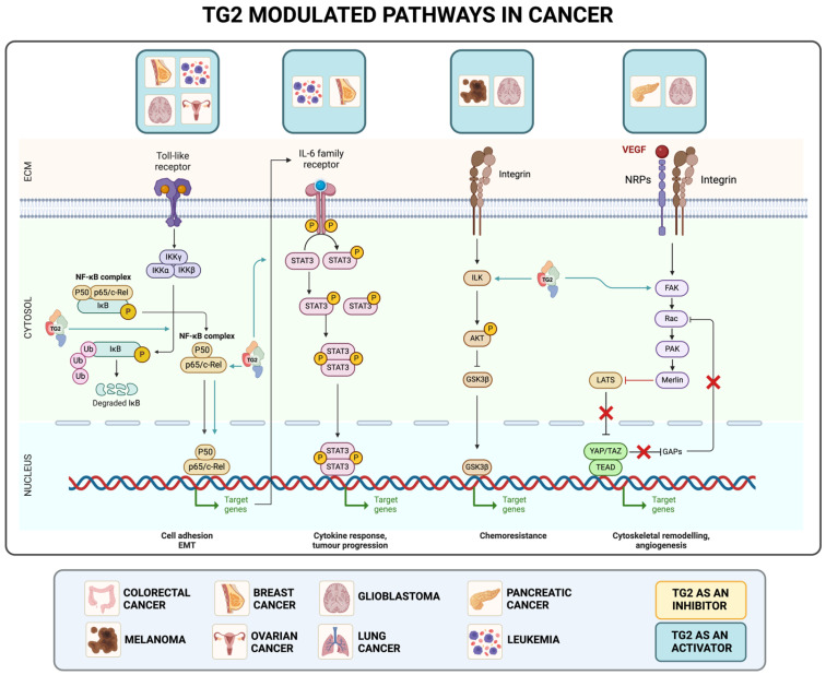
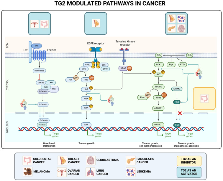
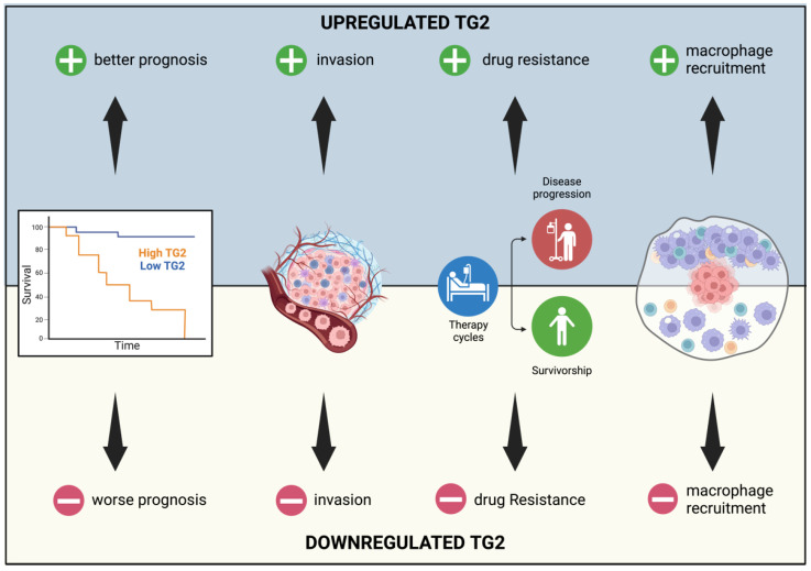
Transglutaminase type 2 (TG2) is the most ubiquitously expressed and well characterized member of the transglutaminase family. It is a ubiquitous multifunctional enzyme implicated in the regulation of several cellular pathways that support the survival, death, and general homeostasis of eukaryotic cells. Due to its multiple localizations both inside and outside the cell, TG2 participates in the regulation of many crucial intracellular signaling cascades in a tissue- and cell-specific manner, making this enzyme an important player in disease development and progression. Moreover, TG2 is capable of modulating the tumor microenvironment, a process of dynamic tissue remodeling and biomechanical events, resulting in changes which influence tumor initiation, growth, and metastasis. Even if generally related to the Ca2+-dependent post-translational modification of proteins, a number of different biological functions have been ascribed to TG2, like those of a peptide isomerase, protein kinase, guanine nucleotide binder, and cytosolic-nuclear translocator. With respect to cancer, TG2's role is controversial and highly debated; it has been described both as an anti- and pro-apoptotic factor and is linked to all the processes of tumorigenesis. However, numerous pieces of evidence support a tissue-specific role of TG2 so that it can assume both oncogenic and tumor-suppressive roles.

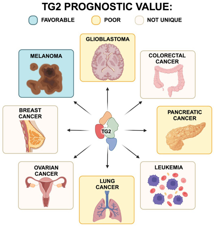
Keywords: breast cancer; colorectal cancer; leukemia; lung cancer; melanoma; microenvironment; ovarian cancer; pancreatic cancer; transglutaminase 2.


Conflict of interest statement

The authors declare no conflicts of interest.

Figures

Figure 1
Figure 1
Representation of TG2′s structure from https://alphafold.ebi.ac.uk, accessed on 21 January 2024. (A) Tertiary structure of TG2. Different colors represent different domains. (B) TG2′s molecular surface. Different colors represent different domains.
Figure 2
Figure 2
TGM2 expression level in tumors vs. normal tissues on GEPIA. SKCM (skin cutaneous melanoma), BRCA (breast invasive carcinoma), GBM (glioblastoma multiforme), OV (ovarian serous cystadenocarcinoma), COAD (colon adenocarcinoma), LAML (acute myeloid leukemia), DLBC (lymphoid neoplasm diffuse large b-cell lymphoma), PAAD (pancreatic adenocarcinoma)0, and LUAD+LUSC (lung adenocarcinoma + lung squamous cell carcinoma). One-way ANOVA; p-value: * = <0.01.
Figure 3
Figure 3
Overall survival based on TGM2 expression level in SKCM (skin cutaneous melanoma), BRCA (breast invasive carcinoma), GBM (glioblastoma multiforme), OV (ovarian serous cystadenocarcinoma), COAD (colon adenocarcinoma), LAML (acute myeloid leukemia), DLBC (lymphoid neoplasm diffuse large b-cell lymphoma), PAAD (pancreatic adenocarcinoma), and LUAD + LUSC (lung adenocarcinoma + lung squamous cell carcinoma) was obtained through a Kaplan–Meier curve by sorting samples for high (yellow line) and low TGM2 (blue line) expression groups according to quartile (high cutoff = 25%; low cutoff = 75%) on GEPIA. Percent survival was plotted, and p-values are shown as per figure specifications. The dotted lines represent the 95% Confidence Interval.
Figure 4
Figure 4
Schematic representation of TG2′s involvement in different pathways and tumors. For each axis, the following are indicated: TG2′s effect (orange TG2: inhibitor; blue TG2: activator), the tumor type in which this signaling is described, and its role in cancer progression. ECM: extracellular matrix.
Figure 4
Figure 4
Schematic representation of TG2′s involvement in different pathways and tumors. For each axis, the following are indicated: TG2′s effect (orange TG2: inhibitor; blue TG2: activator), the tumor type in which this signaling is described, and its role in cancer progression. ECM: extracellular matrix.
Figure 5
Figure 5
Representation of TG2 upregulation and downregulation effects in SKCM. Different levels of TG2 expression modify the characteristics of proliferation, invasiveness, melanin production, and metastasis formation in melanoma cells.
Figure 6
Figure 6
Representation of the effects of TG2 upregulation and downregulation in breast cancer. Different levels of expression of TG2 modify the proliferation capability, the glycolytic metabolism, drug resistance, and the ability to form metastases.
Figure 7
Figure 7
Representation of the effects of TG2 upregulation and downregulation in colorectal cancer. Different levels of expression of TG2 modify proliferation, invasion, angiogenesis, and metastasis capabilities. Moreover, TG2 overexpression correlates with reduced T cell ability to infiltrate and move.
Figure 8
Figure 8
Representation of the effects of TG2 upregulation and downregulation in pancreatic cancer. Different expression levels of TG2 lead to a different prognosis and drug resistance. Moreover, TG2 modulation is correlated with cancer cells’ efficiency at invading tissues and recruiting macrophages.
Figure 9
Figure 9
Prognostic significance of TG2 expression in different types of cancer. Blue: favorable; orange: poor; beige: not unique.

Similar articles

Cited by

References

    1. Tabolacci C., De Martino A., Mischiati C., Feriotto G., Beninati S. The Role of Tissue Transglutaminase in Cancer Cell Initiation, Survival and Progression. Med. Sci. 2019;7:19. doi: 10.3390/medsci7020019. - DOI - PMC - PubMed
    1. Tatsukawa H., Hitomi K. Role of Transglutaminase 2 in Cell Death, Survival, and Fibrosis. Cells. 2021;10:1842. doi: 10.3390/cells10071842. - DOI - PMC - PubMed
    1. Lorand L., Iismaa S.E. Transglutaminase Diseases: From Biochemistry to the Bedside. FASEB J. 2019;33:3–12. doi: 10.1096/fj.201801544R. - DOI - PubMed
    1. Satchwell T.J., Shoemark D.K., Sessions R.B., Toye A.M. Protein 4.2: A Complex Linker. Blood Cells Mol. Dis. 2009;42:201–210. doi: 10.1016/j.bcmd.2009.01.005. - DOI - PubMed
    1. Csobán-Szabó Z., Fésüs L., Király R. Protein-Peptide Based Assay for the Characterization of Human Blood Coagulation Factor XIII-A Isopeptidase Activity: Protein-Based Isopeptidase Assay for FXIII-A. Anal. Biochem. 2020;600:113699. doi: 10.1016/j.ab.2020.113699. - DOI - PubMed

Substances